. 2010 Jul 30;285(31):23629-38.
doi: 10.1074/jbc.M110.108696. Epub 2010 Jun 2.

Roles of platelet STIM1 and Orai1 in glycoprotein VI- and thrombin-dependent procoagulant activity and thrombus formation

Affiliations

Roles of platelet STIM1 and Orai1 in glycoprotein VI- and thrombin-dependent procoagulant activity and thrombus formation

Karen Gilio et al. J Biol Chem. .

Abstract

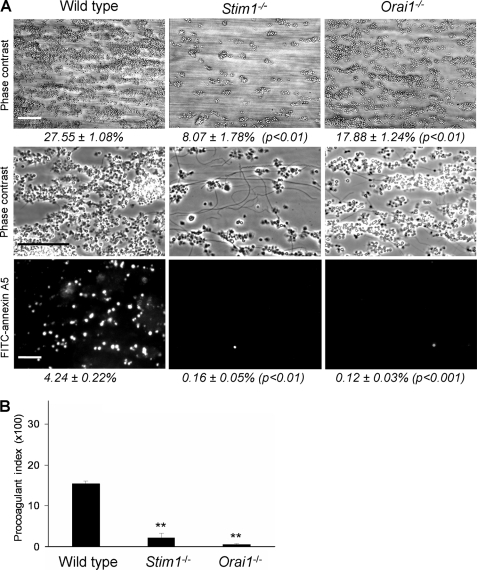
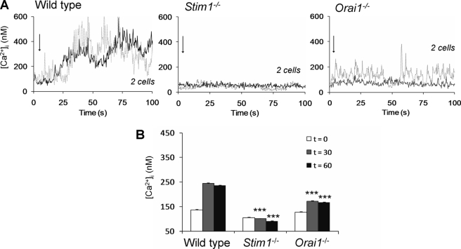
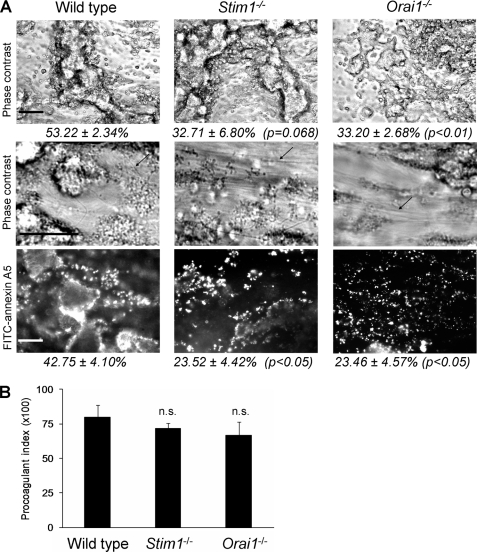
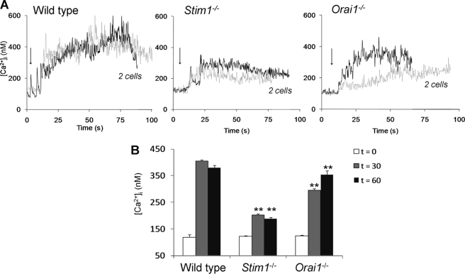
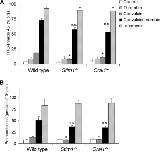
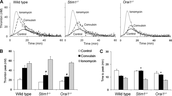
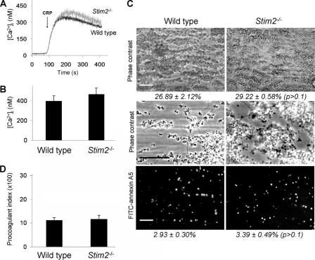
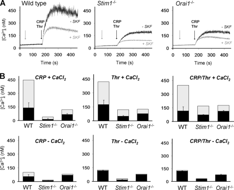
In platelets, STIM1 has been recognized as the key regulatory protein in store-operated Ca(2+) entry (SOCE) with Orai1 as principal Ca(2+) entry channel. Both proteins contribute to collagen-dependent arterial thrombosis in mice in vivo. It is unclear whether STIM2 is involved. A key platelet response relying on Ca(2+) entry is the surface exposure of phosphatidylserine (PS), which accomplishes platelet procoagulant activity. We studied this response in mouse platelets deficient in STIM1, STIM2, or Orai1. Upon high shear flow of blood over collagen, Stim1(-/-) and Orai1(-/-) platelets had greatly impaired glycoprotein (GP) VI-dependent Ca(2+) signals, and they were deficient in PS exposure and thrombus formation. In contrast, Stim2(-/-) platelets reacted normally. Upon blood flow in the presence of thrombin generation and coagulation, Ca(2+) signals of Stim1(-/-) and Orai1(-/-) platelets were partly reduced, whereas the PS exposure and formation of fibrin-rich thrombi were normalized. Washed Stim1(-/-) and Orai1(-/-) platelets were deficient in GPVI-induced PS exposure and prothrombinase activity, but not when thrombin was present as co-agonist. Markedly, SKF96365, a blocker of (receptor-operated) Ca(2+) entry, inhibited Ca(2+) and procoagulant responses even in Stim1(-/-) and Orai1(-/-) platelets. These data show for the first time that: (i) STIM1 and Orai1 jointly contribute to GPVI-induced SOCE, procoagulant activity, and thrombus formation; (ii) a compensating Ca(2+) entry pathway is effective in the additional presence of thrombin; (iii) platelets contain two mechanisms of Ca(2+) entry and PS exposure, only one relying on STIM1-Orai1 interaction.


Figures

FIGURE 1.
FIGURE 1.
Deficiency in STIM1 or Orai1 impedes GPVI-dependent thrombus formation and PS exposure under flow. PPACK/heparin-anticoagulated blood of C57BL/6 mice transplanted with bone marrow from wild type, Stim1−/− or Orai1−/− animals was flowed over collagen at a shear rate of 1000 s−1. A, representative contrast images after 4 min, captured at low (top panels) or high (middle panels) magnification. Bottom panels, fluorescence images after staining with FITC-annexin A5. Percentages in italic indicate area covered with platelets (scale bars, 50 μm). B, procoagulant index representing relative number of PS-exposing platelets. Data are percentage fractions of adhered platelets exposing PS. Means ± S.E. (error bars) are shown. n = 6–8; **, p < 0.01 versus wild type.
FIGURE 2.
FIGURE 2.
Deficiency in STIM1 or Orai1 impedes GPVI-dependent Ca2+ responses of collagen-adhered platelets under flow. PPACK/heparin-anticoagulated blood of wild type or chimeric Stim1−/− or Orai1−/− mice was supplemented with 10% Fluo-4-loaded platelets of the same genotype. Blood samples were flowed over collagen at 1000 s−1, and fluorescence images from the collagen surface were recorded at 5 Hz. A, single-cell rises in Ca2+ of two representative platelets per genotype. Arrows indicate time point of adhesion. B, quantitative analysis of Ca2+ responses at 30 and 60 s after initial Ca2+ rises. Means ± S.E. are shown. n = 35–45 cells; ***, p < 0.001 compared with wild type.
FIGURE 3.
FIGURE 3.
Deficiency in STIM1 or Orai1 permits GPVI-dependent thrombus formation and PS exposure in the presence of coagulation. Citrate-anticoagulated blood of the indicated mice was recalcified with CaCl2/MgCl2 and flowed over collagen for 4 min. Thrombi with platelets and fibrin (arrows) were poststained with FITC-annexin A5. A, representative phase contrast and fluorescence images after 4 min (scale bars, 50 μm). Percentages in italic indicate area covered with (fluorescent) platelets. B, procoagulant index of relative number of PS-exposing platelets. Means ± S.E. (error bars) are shown. n = 5–7; n.s., difference between groups not significant.
FIGURE 4.
FIGURE 4.
Deficiency in STIM1 or Orai1 partly reduces Ca2+ signaling in the presence of coagulation. Citrate-anticoagulated blood of the indicated mice was supplemented with 10% Fluo-4-loaded platelets of the same genotype and recalcified. During blood flow over collagen, fluorescence images from the collagen surface were recorded at 5 Hz. A, single-cell rises in [Ca2+]i of two representative platelets per genotype. Arrows indicate time point of adhesion. B, quantitative analysis of Ca2+ responses at 30 and 60 s after initial [Ca2+]i rises. Means ± S.E. (error bars) are shown. n = 15–29 cells; **, p < 0.01 compared with wild type.
FIGURE 5.
FIGURE 5.
Deficiency in STIM1 or Orai1 permits GPVI-dependent procoagulant activity in the presence of thrombin. Platelets in buffer containing 2 mm CaCl2 (1 × 10⁸/ml) were stimulated for 10 min with convulxin (100 ng/ml), thrombin (4 nm), or ionomycin (20 μm), as indicated. A, flow cytometric analysis of binding of FITC-annexin A5 to platelets (n = 5). B, determination of prothrombinase activity of platelet suspensions. Activity is expressed as pmol of thrombin/min per 106 platelets. n = 7–8. Means ± S.E. (error bars) are shown. *, p < 0.05; n.s., not significant compared with wild type.
FIGURE 6.
FIGURE 6.
Deficiency in STIM1 or Orai1 reduces GPVI-dependent thrombin generation. Citrate-anticoagulated PRP (1 × 10⁸ platelets/ml) of the indicated mice was preincubated with vehicle solvent (control), convulxin (100 ng/ml), or ionomycin (20 μm). Thrombin generation was triggered with tissue factor/CaCl2. A, representative thrombin generation curves per genotype. B, quantification of thrombin peak height. C, quantification of time to peak. Means ± S.E. (error bars) are shown. n = 4–6. #, p < 0.05 compared with wild type (ionomycin); *, p < 0.05 compared with control.
FIGURE 7.
FIGURE 7.
Unchanged GPVI-dependent Ca2+ responses and thrombus formation of STIM2-deficient platelets. A, representative Ca2+ rises of Fura-2-loaded platelets from wild type and Stim2−/− mice, induced by CRP (10 μg/ml) plus CaCl2 (1 mm). B, quantification of maximal Ca2+ rises. C and D, thrombus formation after flow of PPACK/heparin-anticoagulated blood over collagen at high shear rate (4 min). C, representative contrast images captured at low (top panels) or high (middle panels) magnification. Bottom panels, fluorescence images after staining with FITC-annexin A5 (scale bars, 50 μm). D, procoagulant index of relative number of PS-exposing platelets. Means ± S.E. (error bars) are shown. n = 4–6. The difference between groups was not significant.
FIGURE 8.
FIGURE 8.
Residual receptor-induced Ca2+ entry in STIM1- and Orai1-deficient platelets. Calcium responses were measured of Fura-2-loaded platelets from wild type (WT), chimeric Stim1−/−, or Orai1−/− mice, induced by CRP (10 μg/ml) and/or thrombin (Thr, 0.9 nm) and CaCl2 (1 mm), as indicated. A, representative traces of SKF96365 (SKF, 100 μm) effect on Ca2+ rises. Arrows indicate addition of SKF96365 and agonist(s), respectively. B, quantitative effect of SKF96365 on Ca2+ rises. Total bars give rises without SKF96365; black bars indicate rises with SKF96365; and gray bars represent inhibition by SKF96365 (means ± S.E., n = 3–4).

Similar articles

Cited by

References

    1. Heemskerk J. W., Bevers E. M., Lindhout T. (2002) Thromb. Haemost. 88, 186–193 - PubMed
    1. Heemskerk J. W., Feijge M. A., Henneman L., Rosing J., Hemker H. C. (1997) Eur. J. Biochem. 249, 547–555 - PubMed
    1. Zwaal R. F., Schroit A. J. (1997) Blood 89, 1121–1132 - PubMed
    1. Heemskerk J. W., Kuijpers M. J., Munnix I. C., Siljander P. R. (2005) Trends Cardiovasc. Med. 15, 86–92 - PubMed
    1. Munnix I. C., Strehl A., Kuijpers M. J., Auger J. M., van der Meijden P. E., van Zandvoort M. A., oude Egbrink M., Nieswandt B., Heemskerk J. W. (2005) Arterioscler. Thromb. Vasc. Biol. 25, 2673–2678 - PubMed

Publication types