. 2010 Jun 2;30(22):7528-37.
doi: 10.1523/JNEUROSCI.0379-10.2010.

Spinal cord injury immediately changes the state of the brain

Affiliations

Spinal cord injury immediately changes the state of the brain

Juan Aguilar et al. J Neurosci. .

Abstract

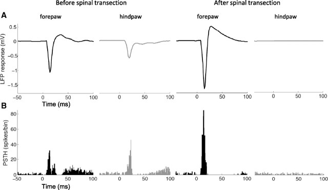
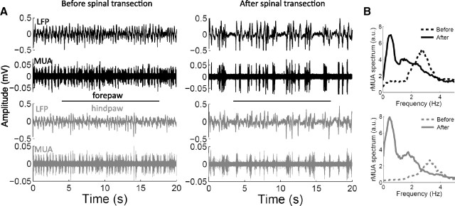
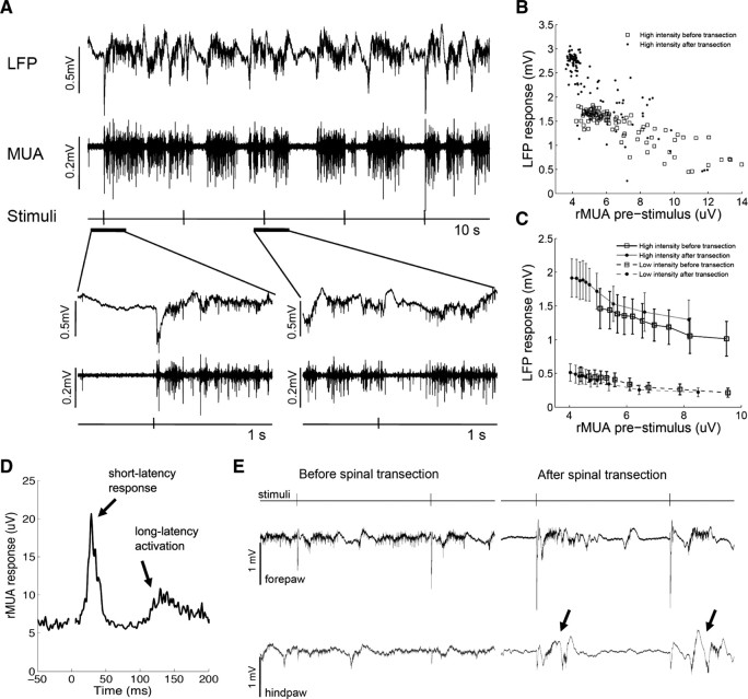
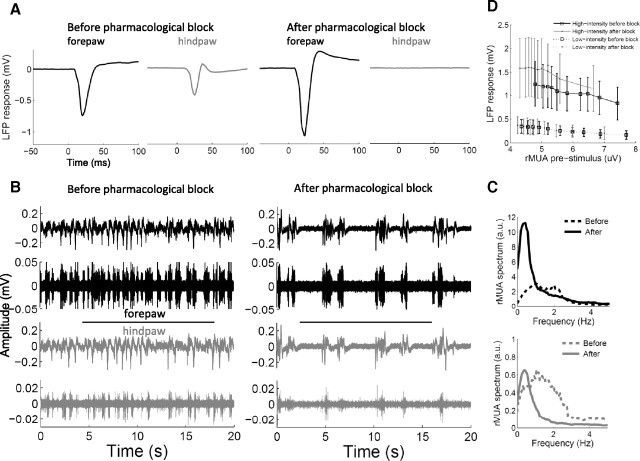
Spinal cord injury can produce extensive long-term reorganization of the cerebral cortex. Little is known, however, about the sequence of cortical events starting immediately after the lesion. Here we show that a complete thoracic transection of the spinal cord produces immediate functional reorganization in the primary somatosensory cortex of anesthetized rats. Besides the obvious loss of cortical responses to hindpaw stimuli (below the level of the lesion), cortical responses evoked by forepaw stimuli (above the level of the lesion) markedly increase. Importantly, these increased responses correlate with a slower and overall more silent cortical spontaneous activity, representing a switch to a network state of slow-wave activity similar to that observed during slow-wave sleep. The same immediate cortical changes are observed after reversible pharmacological block of spinal cord conduction, but not after sham. We conclude that the deafferentation due to spinal cord injury can immediately (within minutes) change the state of large cortical networks, and that this state change plays a critical role in the early cortical reorganization after spinal cord injury.


Figures

Figure 1.
Figure 1.
Changes of cortical responses. A, LFP responses evoked in the forepaw cortex by forepaw stimuli (black) and in the hindpaw cortex by hindpaw stimuli (gray) before (left) and immediately after (right) complete thoracic transection of the spinal cord in a representative animal. B, PSTHs representing multiunit responses recorded from the same electrodes. LFP responses and PSTHs were evaluated from 100 stimuli. Time 0 (x-axis) represents stimulus onset. Spinal cord transection completely abolished the responses evoked by hindpaw stimuli, as expected, but remarkably enhanced the responses evoked by forepaw stimuli.
Figure 2.
Figure 2.
Changes of cortical spontaneous activity. A, Examples of 20 s spontaneous recordings of LFPs and MUA from the forepaw cortex (black) and the hindpaw cortex (gray) before (left) and immediately after (right) complete thoracic transection of the spinal cord in a representative animal. B, Power spectrum of the rMUA corresponding to the recordings shown in A. Before spinal cord transection, the somatosensory cortex of this animal was oscillating at delta frequencies (∼3 Hz), but the oscillation switched to slow-wave activity (∼0.5 Hz) after the spinal cord transection.
Figure 3.
Figure 3.
Relation between cortical spontaneous activity and evoked responses. A, Example of 10 s recordings of LFPs and MUA in response to electrical somatosensory stimuli delivered every 2 s. The time around two representative stimuli is expanded on the bottom: if immediately before the stimulus the cortex is silent (small MUA), then the LFP response is large; if immediately before the stimulus the cortex is active (large MUA), then the LFP response is small. B, Single-trial data from a representative animal: rMUA before stimulus (x-axis) is plotted against the LFP response (y-axis) for 100 high-intensity stimuli delivered before (empty squares) and after (filled dots) complete thoracic transection of the spinal cord. C, Pooled data from all the animals (n = 14). For each animal, stimuli were sorted based on the rMUA before stimulus and LFP responses were averaged in blocks of 10 stimuli, to obtain 10 points per animal, which were then averaged across animals. Error bars indicate 95% confidence intervals. The increased evoked responses correlated with the more silent spontaneous activity after spinal cord transection. D, Average rMUA responses evoked in the hindpaw cortex by high-intensity forepaw stimuli after spinal transection in a representative animal that displayed both short-latency responses and long-latency activations. The signal around time 0 was truncated to eliminate the stimulus artifact. E, Example of LFP recordings throughout two consecutive high-intensity forepaw stimuli (interstimulus interval 2 s) before (left) and after (right) spinal cord transection. During slow-wave activity, high-intensity forepaw stimuli triggered active states in the forepaw cortex that propagated to the hindpaw cortex, generating long-latency activations (arrows).
Figure 4.
Figure 4.
Cortical changes after pharmacological block of the spinal cord. A, Changes of cortical responses in a representative animal (as in Fig. 1A). B, C, Changes in cortical spontaneous activity in a representative animal (as in Fig. 2A,B). D, Relation between cortical spontaneous activity and evoked responses in all animals (n = 8) (as in Fig. 3C). The cortical changes observed after pharmacological block of the spinal cord were very similar to the ones observed after spinal cord transection.

Similar articles

Cited by

References

    1. Aguilar JR, Castro-Alamancos MA. Spatiotemporal gating of sensory inputs in thalamus during quiescent and activated states. J Neurosci. 2005;25:10990–11002. - PMC - PubMed
    1. Aguilar J, Morales-Botello ML, Foffani G. Tactile responses of hindpaw, forepaw and whisker neurons in the thalamic ventrobasal complex of anesthetized rats. Eur J Neurosci. 2008;27:378–387. - PMC - PubMed
    1. Amzica F, Steriade M. Disconnection of intracortical synaptic linkages disrupts synchronization of a slow oscillation. J Neurosci. 1995;15:4658–4677. - PMC - PubMed
    1. Arieli A, Sterkin A, Grinvald A, Aertsen A. Dynamics of ongoing activity: explanation of the large variability in evoked cortical responses. Science. 1996;273:1868–1871. - PubMed
    1. Arthurs OJ, Boniface SJ. What aspect of the fMRI BOLD signal best reflects the underlying electrophysiology in human somatosensory cortex? Clin Neurophysiol. 2003;114:1203–1209. - PubMed

Publication types

MeSH terms