. 2010 Jun;82(6):996-1003.
doi: 10.4269/ajtmh.2010.09-0022.

Isolation and characterization of the MSP1 genes from Plasmodium malariae and Plasmodium ovale

Affiliations

Isolation and characterization of the MSP1 genes from Plasmodium malariae and Plasmodium ovale

Larry Birkenmeyer et al. Am J Trop Med Hyg. 2010 Jun.

Abstract

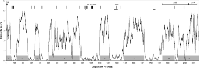
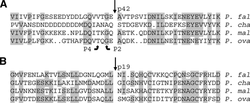
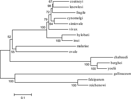
The merozoite surface protein 1 (MSP1) is the principal surface antigen of the blood stage form of the Plasmodium parasite. Antibodies recognizing MSP1 are frequently detected following Plasmodium infection, making this protein a significant component of malaria vaccines and diagnostic tests. Although the MSP1 gene sequence has been reported for Plasmodium falciparum and Plasmodium vivax, this gene has not been identified for the other two major human-infectious species, Plasmodium malariae and Plasmodium ovale. MSP1 genes from these two species were isolated from Cameroon blood donor samples. The genes are similar in size to known MSP1 genes and encode proteins with interspecies conserved domains homologous to those identified in other Plasmodium species. Sequence and phylogenetic analysis of all available Plasmodium MSP1 amino acid sequences clearly shows that the Po and Pm MSP1 sequences are truly unique within the Plasmodium genus and not simply Pf or Pv variants.


Figures

Figure 1.
Figure 1.
Interspecies comparison of MSP1 protein sequences. Full-length deduced MSP1 amino acid sequences from 16 Plasmodium species (Table 2) were aligned and an average amino acid similarity score was determined for successive 10 amino acid segments (alignment available upon request). Plasmodium falciparum, P. vivax, and P. ovale were represented by strains K1, Belem, and OM1A, respectively. Shaded boxes indicate locations of reported conserved sequence blocks. Along the top, positions of differences between deduced MSP1 amino acid sequences from P. ovale strains OM1A and OM1B are denoted by vertical lines. Locations of imperfect repeats (horizontal lines) are indicated for P. malariae (upper lines) and P. ovale (OM1A isolate, lower lines). Signal peptide (SP), p33 and p19 regions are also shown.
Figure 2.
Figure 2.
Predicted sites of primary and secondary merozoite surface protein 1 (MSP1) cleavage. Sequence alignments encompassing the known N-terminal cleavage sites (straight arrows) for (A) MSP1-p42 and (B) MSP1-p19 from Plasmodium falciparum strain T9/94 (P. fal) and Plasmodium chabaudi strain AS (P. cha), and the predicted cleavage sites for the homologous regions from Plasmodium malariae strain MM1A (P. mal) and Plasmodium ovale strain OM1A (P. ova) are shown. Aligned residues conserved in at least 3 of the 4 species are shaded, and the P2 and P4 positions are indicated for MSP1-p42.
Figure 3.
Figure 3.
Alignment of C-terminal p19 regions of merozoite surface protein 1 (MSP1). The deduced amino acid sequences encompassing the predicted MSP1-p19 C-terminal regions (underlined) of the indicated Plasmodium species (denoted by P. and the first 3 letters of the species name) were aligned. P. falA and P. falB refer to Plasmodium falciparum strains K1 and MAD20, respectively, and P. viv refers to the Plasmodium vivax Belem strain. Amino acid differences (lower case letters) vs. the Consensus sequence (upper case letters) are shown. Gaps introduced to maximize alignment (dashes), positions identical to the Consensus sequence (dots), and positions within the Consensus lacking a plurality (X) are indicated. Positions of conserved cysteine residues are shaded. The predicted GPI anchor site (arrow) and the conserved residues (box) surrounding it are indicated.
Figure 4.
Figure 4.
MSP1 phylogenetic tree. The evolutionary relatedness of MSP1 sequences was inferred using the neighbor-joining method. Only the amino acid sequences comprising the interspecies conserved blocks of MSP1 were examined, all regions containing gaps were not considered in the analysis. The evolutionary distances were computed using the JTT matrix-based method and are in the units of the number of amino acid substitutions per site. There were a total of 1,280 positions in the final dataset. The optimal tree is shown with the percentage of replicate trees in which the associated taxa cluster together based on the bootstrap test (1,000 replicates) are shown next to the branches.

References

    1. Korenromp E, Miller J, Nahlen B, Wardlaw T, Young M. World Malaria Report. 2005. http://rbm.who.int/wmr2005/html/toc.htm Available at.
    1. Collins WE, Barnwell JW. Plasmodium knowlesi: finally being recognized. J Infect Dis. 2009;199:1107–1108. - PubMed
    1. Lysenko AJ, Beljaev AE. An analysis of the geographical distribution of Plasmodium ovale. Bull World Health Organ. 1969;40:383–394. - PMC - PubMed
    1. Mueller I, Zimmerman PA, Reeder JC. Plasmodium malariae and Plasmodium ovale–the “bashful” malaria parasites. Trends Parasitol. 2007;23:278–283. - PMC - PubMed
    1. Cox-Singh J, Davis TM, Lee KS, Shamsul SS, Matusop A, Ratnam S, Rahman HA, Conway DJ, Singh B. Plasmodium knowlesi malaria in humans is widely distributed and potentially life threatening. Clin Infect Dis. 2008;46:165–171. - PMC - PubMed

Substances