. 2010 Oct 28;116(17):3278-85.
doi: 10.1182/blood-2008-10-183665. Epub 2010 Jun 2.

BCR-ABL SH3-SH2 domain mutations in chronic myeloid leukemia patients on imatinib

Affiliations

BCR-ABL SH3-SH2 domain mutations in chronic myeloid leukemia patients on imatinib

Daniel W Sherbenou et al. Blood. .

Abstract

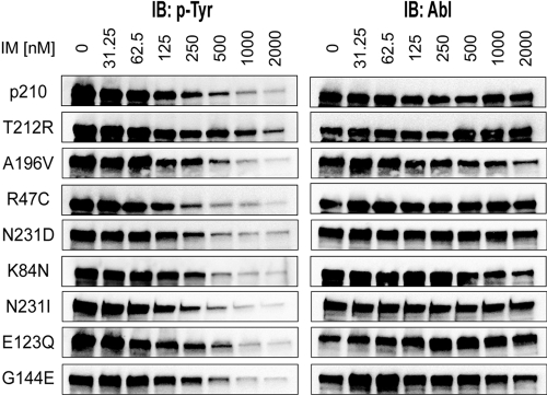
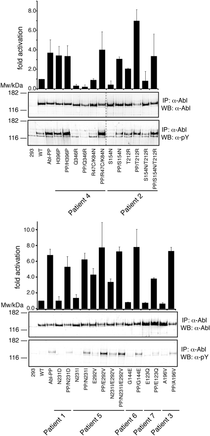
Point mutations in the kinase domain of BCR-ABL are the most common mechanism of drug resistance in chronic myeloid leukemia (CML) patients treated with ABL kinase inhibitors, including imatinib. It has also been shown in vitro that mutations outside the kinase domain in the neighboring linker, SH2, SH3, and Cap domains can confer imatinib resistance. In the context of ABL, these domains have an autoinhibitory effect on kinase activity, and mutations in this region can activate the enzyme. To determine the frequency and relevance to resistance of regulatory domain mutations in CML patients on imatinib, we screened for such mutations in a cohort of consecutive CML patients with various levels of response. Regulatory domain mutations were detected in 7 of 98 patients, whereas kinase domain mutations were detected in 29. One mutation (T212R) conferred in vitro tyrosine kinase inhibitor resistance and was associated with relapse, whereas most other mutations did not affect drug sensitivity. Mechanistic studies showed that T212R increased the activity of ABL and BCR-ABL and that T212R-induced resistance may be partially the result of stabilization of an active kinase conformation. Regulatory domain mutations are uncommon but may explain resistance in some patients without mutations in the kinase domain.


Figures

Figure 1
Figure 1
Follow-up data for patients with detection of a regulatory domain mutation. (A) Available quantitative RT-PCR data for BCR-ABL in patients with a SH3-SH2 domain mutation were graphed for the period of 12 months before to 36 months after the index sample with mutation detection. (B) Clinical timelines corresponding to the quantitative RT-PCR data are shown to indicate the cytogenetic and mutation analysis during the study period. All patients were taking imatinib during the period shown, unless switched to dasatinib when noted.
Figure 2
Figure 2
Locations of patient regulatory domain mutations in the autoinhibited conformations of ABL. The crystal structure of autoinhibited ABL in complex with the kinase inhibitor PD166326 (PDB entry 2FO0) displayed with the locations of the mutated residue side chains shown in green stick format. The mutations are dispersed across the SH2 (green), SH3 (yellow), and linker domains (red, magenta). Stick format atoms are color-coded for nitrogen (dark blue), oxygen (red), and sulfur (orange). The figure was created using PyMol.
Figure 3
Figure 3
Imatinib sensitivity of regulatory domain mutations in BCR-ABL. SH3-SH2 mutations were introduced into BCR-ABL and stably expressed in Ba/F3 cells. These cell lines were exposed to graded concentrations of imatinib for 4 hours and lysed directly into sodium dodecyl sulfate-polyacrylamide gel electrophoresis loading buffer. Immunoblots were performed for BCR-ABL autophosphorylation, and expression. T212R was the only mutant to display detectable resistance of BCR-ABL autophosphorylation to imatinib.
Figure 4
Figure 4
Kinase activation of regulatory domain mutations in ABL. Immunoprecipitated ABL proteins expressing patient SH3-SH2 domain mutations alone or together with the ABL-activating PP (P223E/P230E) mutations were assayed for activity by in vitro kinase assays with an optimal substrate peptide. The graphs show catalytic activity relative to ABL for 4 experiments (from 2 independent transfections) done in duplicate (mean ± SD). Representative immunoblots showing equal Abl protein levels (top panel) and degree of Abl autophosphorylation (bottom panel) are shown below the bar graphs. Note that, in the left panel, 2 lanes with unrelated samples were cropped between lanes 8 and 9 (indicated by dashed line).
Figure 5
Figure 5
Compensatory mutation analysis in the T212R mutant. (A) The threonine of residue 212 (green) is located near the interface between SH2 and the kinase domain's N-terminal lobe in the active conformation of ABL (PDB entry 1OPL, molecule B). (B) In the T212R mutant, arginine is in position to form an electrostatic interaction with E275 in the kinase domain (gray). The kinase domain is shown in gray with bound inhibitor in yellow. Stick format atoms are color-coded for nitrogen (dark blue), oxygen (red), and sulfur (orange). The figure was created using PyMol. (C) Single-, double-, and triple-mutant ABL expression constructs with T212R, ABL PP (P223E/P230E), and mutations at the E275 position were transiently transfected in HEK293 cells, lysed 40 hours later, and total protein extracts analyzed by anti-ABL and antiphosphotyrosine immunoblotting. (D) These ABL mutant proteins were next immunoprecipitated and assayed for activity by in vitro kinase assays with an optimal substrate peptide. The graphs show catalytic activity relative to ABL for 2 experiments done in duplicate (mean ± SD). (E) The E275K and S154N mutants were tested in native and T212R BCR-ABL by Ba/F3 cell proliferation assays, as before. E275K displayed substantial imatinib resistance alone, and this effect was additive to that of T212R in the double mutants. T212R/E275A had the same sensitivity to that of the T212R single mutant. In the T212R/S154N double mutant, the sensitivity of imatinib was reverted back to that of WT BCR-ABL.
Figure 6
Figure 6
Kinase activity of full-length T212R BCR-ABL. Native BCR-ABL and T212R mutant BCR-ABL were overexpressed in HEK293 cells and immunoprecipitated in triplicate. Kinase assays were performed in triplicate at the indicated substrate concentrations, and the enzyme velocity was plotted as a function of peptide concentration. BCR-ABL levels were quantified from anti-ABL immunoblots of immunoprecipitated native and T212R mutant BCR-ABL and used to normalize levels of protein used in the kinase assay.

Similar articles

Cited by

References

    1. Druker BJ, Guilhot F, O'Brien SG, et al. Five-year follow-up of patients receiving imatinib for chronic myeloid leukemia. N Engl J Med. 2006;355(23):2408–2417. - PubMed
    1. Cortes J, O'Brien S, Kantarjian H. Discontinuation of imatinib therapy after achieving a molecular response. Blood. 2004;104(7):2204–2205. - PubMed
    1. Higashi T, Tsukada J, Kato C, et al. Imatinib mesylate-sensitive blast crisis immediately after discontinuation of imatinib mesylate therapy in chronic myelogenous leukemia: report of two cases. Am J Hematol. 2004;76(3):275–278. - PubMed
    1. Sawyers CL, Hochhaus A, Feldman E, et al. Imatinib induces hematologic and cytogenetic responses in patients with chronic myelogenous leukemia in myeloid blast crisis: results of a phase II study. Blood. 2002;99(10):3530–3539. - PubMed
    1. Branford S, Rudzki Z, Walsh S, et al. High frequency of point mutations clustered within the adenosine triphosphate-binding region of BCR/ABL in patients with chronic myeloid leukemia or Ph-positive acute lymphoblastic leukemia who develop imatinib (STI571) resistance. Blood. 2002;99(9):3472–3475. - PubMed

Publication types

MeSH terms