. 2010 Sep 2;116(9):1506-14.
doi: 10.1182/blood-2010-01-265686. Epub 2010 Jun 2.

microRNA-dependent modulation of histone acetylation in Waldenstrom macroglobulinemia

Affiliations

microRNA-dependent modulation of histone acetylation in Waldenstrom macroglobulinemia

Aldo M Roccaro et al. Blood. .

Abstract

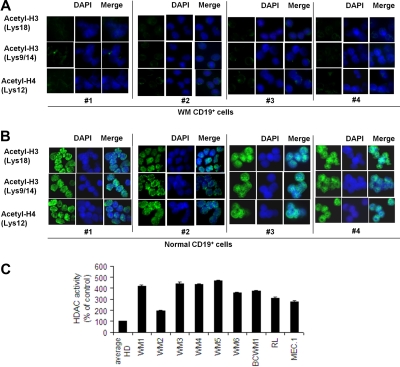
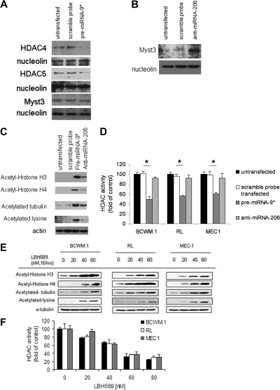
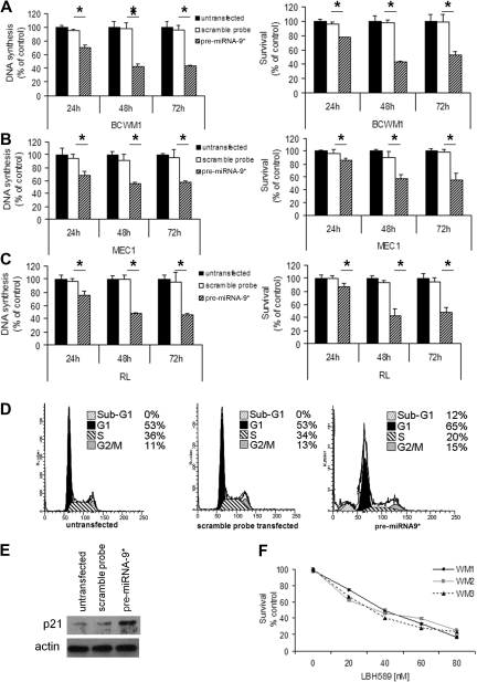
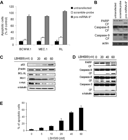
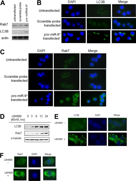
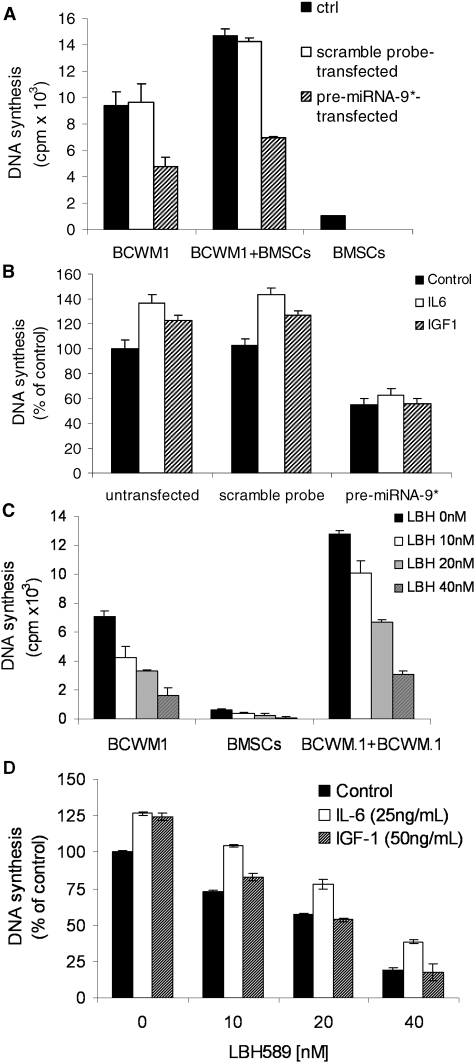
Waldenström macroglobulinemia (WM) cells present with increased expression of microRNA-206 (miRNA-206) and reduced expression of miRNA-9*. Predicted miRNA-206- and -9*-targeted genes include histone deacetylases (HDACs) and histone acetyl transferases (HATs), indicating that these miRNAs may play a role in regulating histone acetylation. We were able to demonstrate that primary WM cells are characterized by unbalanced expression of HDACs and HATs, responsible for decreased acetylated histone-H3 and -H4, and increased HDAC activity. We next examined whether miRNA-206 and -9* modulate the aberrant expression of HDAC and HATs in WM cells leading to increased transcriptional activity. We found that restoring miRNA-9* levels induced toxicity in WM cells, supported by down-modulation of HDAC4 and HDAC5 and up-regulation of acetyl-histone-H3 and -H4. These, together with inhibited HDAC activity, led to induction of apoptosis and autophagy in WM cells. To further confirm that miRNA-9*-dependent modulation of histone acetylation is responsible for induction of WM cytotoxicity, a novel class of HDAC inhibitor (LBH589) was used; we confirmed that inhibition of HDAC activity leads to toxicity in this disease. These findings confirm that histone-modifying genes and HDAC activity are deregulated in WM cells, partially driven by the aberrant expression of miRNA-206 and -9* in the tumor clone.


Figures

Figure 1
Figure 1
Primary WM cells present with higher level of miRNA-206, lower level of miRNA-9*, together with increased expression of HDACs and reduced expression of HATs, compared with healthy donors. (A) miRNA analysis has been performed on total RNA isolated from BM CD19+ WM cells, normal bone marrow (NBM)– and PBMC-derived CD19+ counterparts, and WM cell line (BCWM.1). Heatmap was generated after supervised hierarchical clustering analysis was performed with analysis of variance test. Differential expression of miRNA patterns is shown by the intensity of red (up-regulation) versus blue (down-regulation). (B) Purified cRNA (15 μg) isolated from primary CD19+ cells isolated from BM of 6 patients with WM and from CD19+ cells isolated from PBMCs of 2 healthy donors was hybridized to HG-U133Plus2.0 GeneChip (Affimetrix). Fold change is shown by the intensity of induction (red) or suppression (blue).
Figure 2
Figure 2
Lower expression of acetylated histone-3/histone-4 and higher HDAC activity characterize primary WM cells compared with healthy donors. Immunocytochemical analysis of primary CD19+ cells isolated from BM of 4 patients with WM (A) and CD19+ cells isolated from PBMCs of 4 healthy donors (B) was performed with the use of anti–acetyl-histone-H3 (Lys18), -H3 (lys9/14), and -H4 (lys12) antibodies. DAPI (4′-6′-diamidine-2-phenylindole) was used to stain nuclei. (C) HDAC activity was assessed with nuclear extracts with the use of a Colorimetric HDAC Activity Assay Kit on primary CD19+ cells isolated from BM of 6 patients with WM (WM1, WM2, WM3, WM4, WM5, WM6); primary CD19+ cells were isolated from PBMCs of 3 healthy donors (average HD), and BCWM.1 and low-grade lymphomas IgM-secreting cell lines (all P ≤ .05).
Figure 3
Figure 3
miRNA-9* and miRNA-206 target HDAC4/HDAC5 and Myst3, respectively. (A) BCWM.1 cells (scramble probe–, pre-miRNA-9*–transfected, and untransfected) were harvested at 12 hours after transfection. Nuclear lysates were subjected to Western blot with the use of anti-HDAC4, -HDAC5, -Myst3, and -nucleolin antibodies. (B) BCWM.1 cells (scramble probe–, anti–miRNA-206–transfected, and untransfected) were harvested at 12 hours after transfection. Nuclear lysates were subjected to Western blot with the use of anti-Myst3 and -nucleolin antibodies. (C) BCWM.1 cells (scramble probe–, pre-miRNA-9*–, anti–miRNA-206–transfected, and untransfected) were harvested at 12 hours after transfection. Whole-cell lysates were subjected to Western blotting with the use of anti–acetyl-histone H3, –acetyl-histone H4, –acetylated-tubulin, –acetylated-lysine, and -actin antibodies. (D) BCWM.1 cells (scramble probe–, pre-miRNA-9*–, anti–miRNA-206–transfected, and untransfected) were harvested at 12 hours after transfection. HDAC activity was assessed in vitro with the use of nuclear extracts by Colorimetric HDAC Activity Assay Kit (*P < .05). (E) BCWM.1, RL, and MEC-1 cells were cultured with LBH589 (20-60nM) for 16 hours or with control medium. Whole-cell lysates were subjected to Western blotting with the use of anti–acetyl-histone H3, –acetyl-histone H4, –acetylated-tubulin, –acetylated-lysine, and –α-tubulin antibodies. (F) BCWM.1 cells were cultured in the presence or absence of LBH589 (0-80nM; 8 hours). HDAC activity was assessed in vitro with the use of nuclear extracts by Colorimetric HDAC Activity Assay Kit (all P ≤ .05).
Figure 4
Figure 4
miRNA-9* regulates proliferation and survival in WM cells as well as in low-grade lymphoma IgM-secreting cells. (A) WM cells (A: BCWM.1), low-grade lymphoma IgM-secreting cell lines (B: MEC1; C: RL; scramble probe–, precursor (pre)–miRNA-9*–transfected, and untransfected) were harvested at 24, 48, and 72 hours after transfection; DNA synthesis and cytotoxicity were assessed by thymidine uptake and 3-(4,5-dimethylthiazol-2-yl)-2,5-diphenyltetrazolium bromide (MTT) assays, respectively (*P < .05). (D) Cell cycle analysis was performed by propidium iodide staining with BCWM.1 cells (untransfected, scramble probe–, and pre-miRNA-9*–transfected). (E) BCWM.1 cells (untransfected, scramble probe–, and pre-miRNA-9*–transfected) were harvested at 12 hours after transfection. Whole-cell lysates were subjected to Western blotting with anti-p21 and -actin antibodies. (F) Freshly isolated normal CD19+ cells from normal PBMCs were cultured with LBH589 (0-80nM) for 48 hours. Cytotoxicity was assessed by MTS assay.
Figure 5
Figure 5
miRNA-9*–dependent HDAC inhibition exerts a proapoptotic effect on WM cells. (A) Percentage of cells undergoing apoptosis was studied by Apo2.7 staining and flow cytometry in WM and low-grade lymphoma IgM-secreting cell lines (untransfected, scramble probe–, pre-miRNA-9*–transfected were harvested 48 hours after transfection). All P ≤ .05. (B) BCWM.1 cells (untransfected, scramble probe–, and pre-miRNA-9*–transfected) were harvested at 12 hours after transfection. Whole-cell lysates were subjected to Western blot with the use of anti-PARP, –caspase-8, –caspase-9, and –actin antibodies. (C-D) BCWM.1 cells were cultured with LBH589 (0-60nM) for 16 hours. Whole-cell lysates were subjected to Western blot with the use of anti-p53, –BCL-XL, –Mcl-1, –c-myc, –β-actin, -PARP, –caspase-9, –caspase-8, –caspase-3, and –α-tubulin antibodies. (E) BCWM.1 cells were cultured with LBH589 for 48 hours at doses that range from 0 to 60nM, and the percentage of cells undergoing apoptosis was studied by Apo2.7 staining by flow cytometry. All P ≤ .05.
Figure 6
Figure 6
miRNA-9*–dependent HDAC inhibition and modulation of autophagy in WM cells. (A) BCWM.1 cells (untransfected, scramble probe–,and pre-miRNA-9*–transfected) were harvested at 24 hours after transfection. Whole-cell lysates were subjected to Western blot with the use of anti-Rab7, -LC3, and -actin antibodies. (B) BCWM.1 cells were cultured with LBH589 (40nM) for 0 to 24 hours. Whole-cell lysates were subjected to Western blotting with the use of anti-Rab7 and -LC3B antibodies. (C-D) BCWM.1 cells were cultured in the presence or absence of LBH589 (40nM) for 16 hours. Immunocytochemical analysis was assessed with the use of anti-Rab7 (C) or -LC3B (D) antibodies. DAPI indicates 4′-6′-diamidine-2-phenylindole.
Figure 7
Figure 7
miRNA-9*–dependent modulation of WM cell proliferation in the context of BM milieu. (A) BCWM.1 cells (untransfected, scramble probe–, pre-miRNA-9*–transfected) were cultured for 48 hours in the presence or absence of primary BM stromal cells (BMSCs). Cell proliferation was assessed with the use of the [3H]-thymidine uptake assay. (B) BCWM.1 cells (untransfected, scramble probe–, pre-miRNA-9*–transfected) were cultured in the presence or absence of IL-6 (25 ng/mL) or IGF-1 (50 ng/mL) for 48 hours. DNA synthesis was assessed with the use of the [3H]-thymidine uptake assay. (C) BCWM.1 cells were cultured with LBH589 (0-40nM) for 48 hours in the presence or absence of WM patient-derived BMSCs. Cell proliferation was assessed with the use of the [3H]-thymidine uptake assay. (D) BCWM.1 cells were cultured with LBH589 (0-40nM) in the absence and presence of IL-6 (25 ng/mL) or IGF-1 (50 ng/mL) for 48 hours. DNA synthesis was assessed with the use of the [3H]-thymidine uptake assay. All P ≤ .05.

Similar articles

Cited by

References

    1. Ghobrial IM, Gertz MA, Fonseca R. Waldenstrom macroglobulinaemia. Lancet Oncol. 2003;4(11):679–685. - PubMed
    1. Owen RG, Treon SP, Al-Katib A, et al. Clinicopathological definition of Waldenström's macroglobulinemia: consensus panel recommendations from the Second International Workshop on Waldenström's Macroglobulinemia. Semin Oncol. 2003;30(2):110–115. - PubMed
    1. Schop RF, Kuehl WM, Van Wier SA, et al. Waldenström macroglobulinemia neoplastic cells lack immunoglobulin heavy chain locus translocations but have frequent 6q deletions. Blood. 2002;100(8):2996–3001. - PubMed
    1. Chng WJ, Schop RF, Price-Troska T, et al. Gene expression profiling of Waldenström's macroglobulinemia reveals a phenotype more similar to chronic lymphocytic leukemia than multiple myeloma. Blood. 2006;108(8):2755–2763. - PMC - PubMed
    1. Esteller M. Epigenetics in Cancer. N Engl J Med. 2008;358(11):1148–1159. - PubMed

Publication types

MeSH terms