. 2010 Aug;153(4):1795-807.
doi: 10.1104/pp.110.158360. Epub 2010 Jun 2.

Herbivore-induced SABATH methyltransferases of maize that methylate anthranilic acid using s-adenosyl-L-methionine

Affiliations

Herbivore-induced SABATH methyltransferases of maize that methylate anthranilic acid using s-adenosyl-L-methionine

Tobias G Köllner et al. Plant Physiol. 2010 Aug.

Abstract

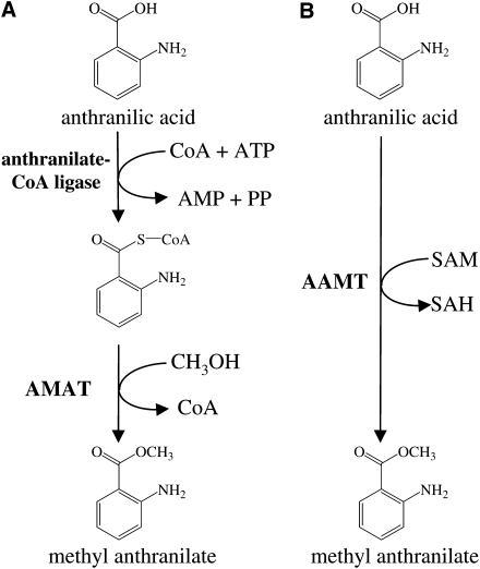
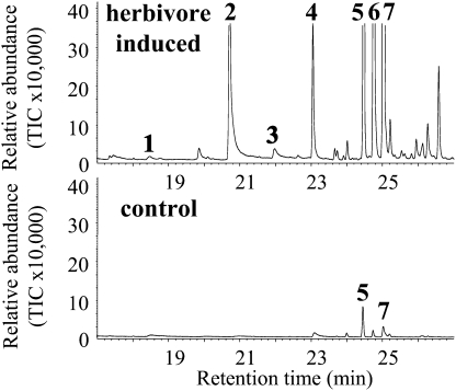
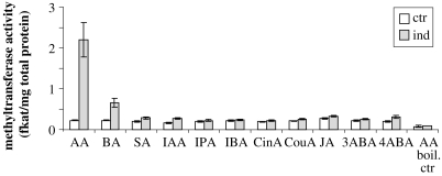
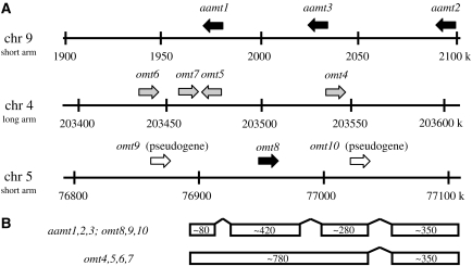
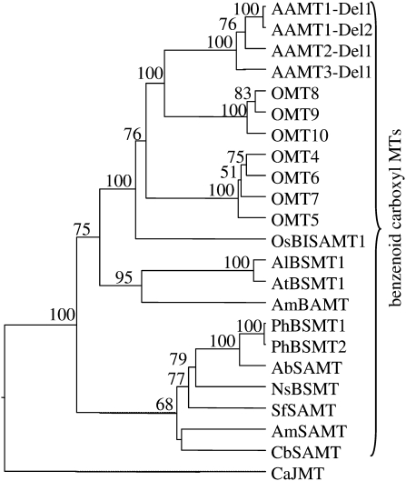
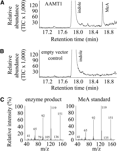
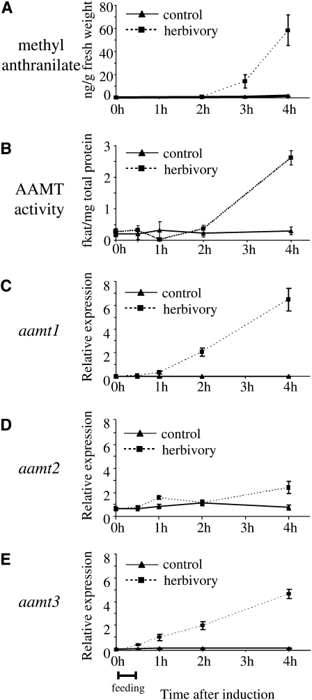
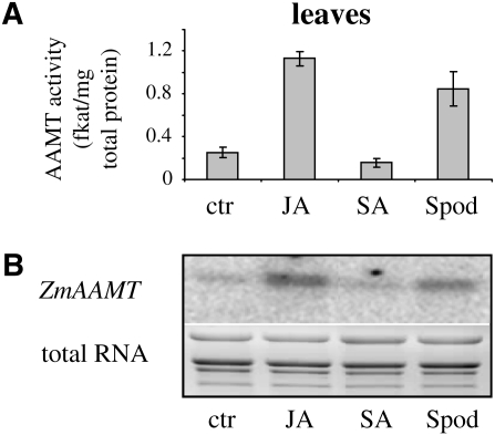
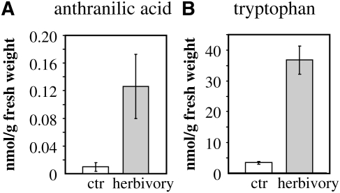
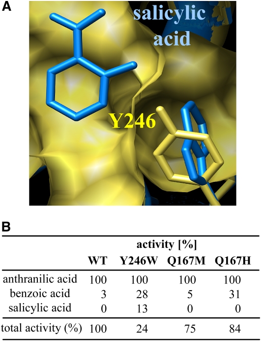
Volatile methyl esters are common constituents of plant volatiles with important functions in plant defense. To study the biosynthesis of these compounds, especially methyl anthranilate and methyl salicylate, we identified a group of methyltransferases that are members of the SABATH enzyme family in maize (Zea mays). In vitro biochemical characterization after bacterial expression revealed three S-adenosyl-L-methionine-dependent methyltransferases with high specificity for anthranilic acid as a substrate. Of these three proteins, Anthranilic Acid Methyltransferase1 (AAMT1) appears to be responsible for most of the S-adenosyl-L-methionine-dependent methyltransferase activity and methyl anthranilate formation observed in maize after herbivore damage. The enzymes may also be involved in the formation of low amounts of methyl salicylate, which are emitted from herbivore-damaged maize. Homology-based structural modeling combined with site-directed mutagenesis identified two amino acid residues, designated tyrosine-246 and glutamine-167 in AAMT1, which are responsible for the high specificity of AAMTs toward anthranilic acid. These residues are conserved in each of the three main clades of the SABATH family, indicating that the carboxyl methyltransferases are functionally separated by these clades. In maize, this gene family has diversified especially toward benzenoid carboxyl methyltransferases that accept anthranilic acid and benzoic acid.


Figures

Figure 1.
Figure 1.
The biosynthesis of methyl anthranilate from anthranilic acid can proceed over two pathways. Pathway A has been documented in grape, while pathway B is demonstrated here. AMAT, Anthraniloyl-CoA:methanol acyltransferase; SAH, S-adenosyl-l-homocysteine.
Figure 2.
Figure 2.
Volatiles released by maize after herbivore attack. Volatiles from 2-week-old plants of maize cv Delprim were collected after no treatment (control) or after 16 h of feeding by Egyptian cotton leaf worm (herbivore induced). After separation by GC, the peaks were identified by MS as methyl salicylate (peak 1), indole (peak 2), methyl anthranilate (peak 3), geranyl acetate (peak 4), (E)-β-caryophyllene (peak 5), (E)-α-bergamotene (peak 6), and (E)-β-farnesene (peak 7). TIC, Total ion current.
Figure 3.
Figure 3.
SAM-dependent methyltransferase activity in undamaged (ctr) and herbivore-damaged (ind) maize seedlings. Plant crude protein extracts were incubated with the methyl group donor [14C]SAM and different acid substrates. The formed methyl esters were extracted from the assays and quantified using a scintillation counter. AA, Anthranilic acid; BA, benzoic acid; SA, salicylic acid; IAA, indole-3-acetic acid; IPA, indole-3-propionic acid; IBA, indole-3-butyric acid; CinA, cinnamic acid; CouA, o-coumaric acid; JA, jasmonic acid; 3ABA, 3-aminobenzoic acid; 4ABA, 4-aminobenzoic acid; boil. ctr, control with heat-denatured enzyme.
Figure 4.
Figure 4.
Chromosomal locations of the benzenoid carboxyl methyltransferase gene family in the maize genome. A, Sections of the maize chromosomes with approximate locations of the SABATH-like genes and pseudogenes. Black arrows represent genes expressed in maize cv Delprim after herbivory; gray arrows represent genes without detectable expression; white arrows represent pseudogenes. B, Exon-intron structure of maize SABATH-like genes.
Figure 5.
Figure 5.
Dendrogram analysis of maize AAMT1-Del1, AAMT1-Del2, AAMT2-Del1, AAMT3-Del1, OMT4, OMT5, OMT6, OMT7, OMT8, OMT9, and OMT10 with different methyltransferases (MTs) specific for carboxyl groups of benzenoids and jasmonic acid. The analysis was conducted using a neighbor-joining algorithm. Bootstrap values are shown in percentage and were generated with a sample of 1,000. Accession numbers are as follows: PhBSMT1, AAO45012; PhBSMT2, AAO45013; AbSAMT, BAB39396; NsBSMT, CAF31508; SfSAMT, CAC33768; AmSAMT, AAN40745; CbSAMT, AAF00108; AlBSMT1, AAP57211; AtBSMT1, NP_187755; AmBAMT, AAF98284; CaJMT, ABB02661; OsBISAMT1, AAS18419. Sequences of OMT4, OMT5, OMT6, OMT7, OMT9, and OMT10 were obtained from the Maize Genome Database (www.maizesequence.org) with the following accession numbers: OMT4, GRMZM2G143871; OMT5, GRMZM2G133996; OMT6, GRMZM2G050307; OMT7, GRMZM2G050321; OMT9, GRMZM2G303419; OMT10, GRMZM2G405947. The ORFs of OMT9 and OMT10 were reconstructed in silico by the deletion of a stop codon (OMT9) or the deletion of two frame-shift mutations (OMT10).
Figure 6.
Figure 6.
GC-MS analysis of methyl anthranilate (MeA) produced by AAMT1. A, The enzyme was expressed in E. coli, extracted, partially purified, and incubated with the substrates SAM and anthranilic acid. The resulting methyl ester was collected with a solid-phase microextraction (SPME) fiber and analyzed by GC-MS. B, GC trace resulting from an empty-vector control experiment. A crude protein extract from E. coli carrying the empty expression vector was incubated with the substrates SAM and AA and analyzed as described above. TIC, Total ion current. C, Comparison of the mass spectrum from the enzyme-produced methyl anthranilate and the mass spectrum derived from an authentic methyl anthranilate standard.
Figure 7.
Figure 7.
Expression analysis of aamt genes in comparison with methyl anthranilate accumulation and AAMT activity after herbivore damage. Plants were treated with S. littoralis larvae caged for 30 min on a single leaf. Leaf material was harvested at different time points after feeding and was split into equal parts for the different analyses. Means and se are shown (n = 4). A, Methyl anthranilate accumulation after herbivore damage in maize leaves. Leaf material was extracted with hexane, and the extracts were analyzed using GC-MS. B, AAMT activity in herbivore-damaged maize leaves. Plant crude protein extracts were incubated with the methyl group donor [14C]SAM and anthranilic acid. The formed methyl ester was extracted from the assays and quantified using a scintillation counter. C to E, Transcript accumulation of aamt1 (C), aamt2 (D), and aamt3 (E) after herbivore damage. Expression was analyzed using quantitative PCR. The relative expression levels were calculated as the expression levels of the respective genes divided by the geometric mean of the expression levels of the two reference genes (for details, see “Materials and Methods”).
Figure 8.
Figure 8.
AAMT activity correlates with transcript accumulation of aamt genes after different plant treatments. Plants were treated with jasmonic acid (JA), salicylic acid (SA), or S. littoralis larvae (Spod). Leaf material was harvested and split into two parts for different analyses. A, AAMT activity. Plant crude protein extracts were incubated with the methyl group donor [14C]SAM and anthranilic acid. The formed methyl ester was extracted from the assays and quantified using a scintillation counter. Means and se are shown (n = 4). B, Transcript accumulation of aamt genes. RNA isolated from leaf material was hybridized with a probe specific for maize aamt genes. The bottom panel shows the total RNA of the ethidium bromide-stained RNA gel. ctr, Control plants without treatment.
Figure 9.
Figure 9.
Levels of anthranilic acid (A) and Trp (B) in herbivore-damaged maize plants (herbivory) in comparison with undamaged control plants (ctr). Anthranilic acid was extracted with methanol from leaf material and analyzed using LC-MS. Trp present in a crude leaf extract was derivatized with mercaptoethanol and O-phthaldialdehyde and analyzed by HPLC coupled to a fluorescence detector. Means and se are shown (n = 5).
Figure 10.
Figure 10.
A, Model of part of the AAMT1 active site cavity based on the structure of C. brewerii SAMT (Zubieta et al., 2003) with the proposed position of salicylic acid. The surface of the active site cavity and the residue Tyr-246 of AAMT1 are shown in yellow. The structure of CbSAMT was overlaid, but only the bound substrate salicylic acid and the residue Trp-226 were visualized (blue). B, Relative activity of wild-type AAMT1 (WT) in comparison with several AAMT1 mutant enzymes with various substrates. Values are averages of four independent measurements. All substrates were tested at a 1.5 mm concentration, and methyltransferase activity was determined by measuring the radioactivity of the transferred 14C-methyl group from SAM. The relative activity of the three AAMTs with anthranilic acid was set arbitrarily at 100%.

Similar articles

Cited by

References

    1. Barkman TJ, Martins TR, Sutton E, Stout JT. (2007) Positive selection for single amino acid change promotes substrate discrimination of a plant volatile-producing enzyme. Mol Biol Evol 24: 1320–1329 - PubMed
    1. Cardoza YJ, Tumlinson JH. (2006) Compatible and incompatible Xanthomonas infections differentially affect herbivore-induced volatile emission by pepper plants. J Chem Ecol 32: 1755–1768 - PubMed
    1. Chen F, D'Auria JC, Tholl D, Ross JR, Gershenzon J, Noel JP, Pichersky E. (2003) An Arabidopsis thaliana gene for methylsalicylate biosynthesis, identified by a biochemical genomics approach, has a role in defense. Plant J 36: 577–588 - PubMed
    1. D'Alessandro M, Held M, Triponez Y, Turlings TCJ. (2006) The role of indole and other shikimic acid derived maize volatiles in the attraction of two parasitic wasps. J Chem Ecol 32: 2733–2748 - PubMed
    1. D'Auria JC, Chen F, Pichersky E. (2003) The SABATH family of MTS in Arabidopsis thaliana and other plant species. Romeo JT, , Integrative Phytochemistry: From Ethnobotany to Molecular Ecology. Recent Advances in Phytochemistry, Vol 37 Elsevier, Oxford, pp 253–283

Publication types

LinkOut - more resources