. 2010 Jun;176(6):2658-68.
doi: 10.2353/ajpath.2010.090690.

Cardiotoxicity of the anticancer therapeutic agent bortezomib

Affiliations

Cardiotoxicity of the anticancer therapeutic agent bortezomib

Dominika Nowis et al. Am J Pathol. 2010 Jun.

Abstract

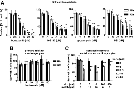
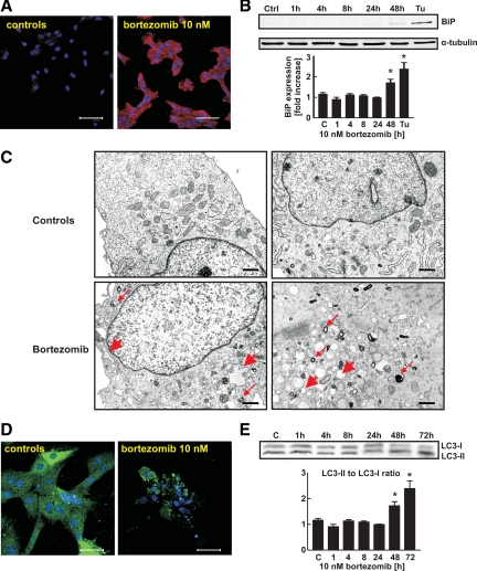
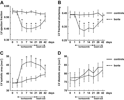
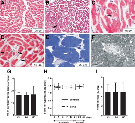
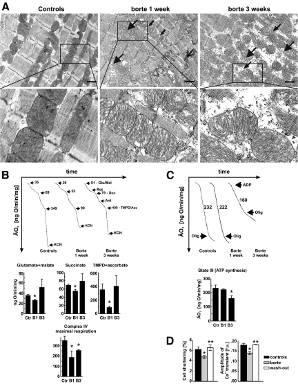
Recent case reports provided alarming signals that treatment with bortezomib might be associated with cardiac events. In all reported cases, patients experiencing cardiac problems were previously or concomitantly treated with other chemotherapeutics including cardiotoxic anthracyclines. Therefore, it is difficult to distinguish which components of the therapeutic regimens contribute to cardiotoxicity. Here, we addressed the influence of bortezomib on cardiac function in rats that were not treated with other drugs. Rats were treated with bortezomib at a dose of 0.2 mg/kg thrice weekly. Echocardiography, histopathology, and electron microscopy were used to evaluate cardiac function and structural changes. Respiration of the rat heart mitochondria was measured polarographically. Cell culture experiments were used to determine the influence of bortezomib on cardiomyocyte survival, contractility, Ca(2+) fluxes, induction of endoplasmic reticulum stress, and autophagy. Our findings indicate that bortezomib treatment leads to left ventricular contractile dysfunction manifested by a significant drop in left ventricle ejection fraction. Dramatic ultrastructural abnormalities of cardiomyocytes, especially within mitochondria, were accompanied by decreased ATP synthesis and decreased cardiomyocyte contractility. Monitoring of cardiac function in bortezomib-treated patients should be implemented to evaluate how frequently cardiotoxicity develops especially in patients with pre-existing cardiac conditions, as well as when using additional cardiotoxic drugs.


Figures

Figure 1
Figure 1
Proteasome inhibitors induce cytostatic/cytotoxic effects in H9c2 rat cardiac myoblast cells. A: H9c2 cells were incubated for 48 or 72 hours with indicated concentrations of selected proteasome inhibitors: bortezomib, MG132, epoxomycin, and carbobenzoxyl-lle-Glu-(O-t-butyl)-Ala-Leucinal (PSI). The cytostatic/cytotoxic effects were measured with crystal violet staining. B: Primary ventricular cardiomyocytes isolated from adult rats were incubated with bortezomib. The cytostatic/cytotoxic effects were measured with crystal violet staining. C: Contractile neonatal ventricular rat cardiomyocytes were incubated with bortezomib. The cytostatic/cytotoxic effects were measured with XTT assay. Bars represent percent survival versus untreated controls. Data are mean ± SD *P < 0.05 versus controls (two-tailed Student’s t-test). **P < 0.05 versus chemotherapy alone (two-tailed Student’s t-test).
Figure 2
Figure 2
Bortezomib induces ER stress and autophagy in H9c2 cells.A: H9c2 cells were incubated for 24 hours with 10 nmol/L bortezomib and subjected to indirect immunofluorescence microscopy using anti-FK2 (multiubiquitin) antibody (red) and 4,6-diamidino-2-phenylindole staining (blue). Scale bars = 75 μm. B: H9c2 cells were incubated with 10 nmol/L bortezomib for indicated periods. Total cell lysates were prepared and Western blot analysis was performed using anti-BiP and anti-α tubulin antibodies. Cells incubated for 16 hours with 10 μg/ml tunicamycin were used as a positive control. Data are mean ± SE *P < 0.05 versus controls (two-tailed Student’s t-test). C: H9c2 cells were incubated for 48 hours with 10 nmol/L bortezomib, collected, and fixed as described in Materials and Methods, and observed by TEM. Thin red arrows indicate multilamellar and/or lysosomal/autophagosomal structures; thick red arrows point to widened endoplasmic reticulum. Scale bars = 200 nm. D: H9c2 cells were transiently transfected with pEGFP-LC3m plasmid encoding EGFP-LC3 fusion protein, incubated for 48 hours with 10 nmol/L bortezomib, fixed, stained with anti-GFP antibody and TO-PRO nuclear stain and observed under fluorescence microscopy. Scale bars = 75 μm. E: H9c2 cells were incubated with 10 nmol/L bortezomib for the indicated periods. Then total cell lysates were prepared and Western blot analysis was performed using anti-LC3B antibody against LC3B-I and LC3B-II fragments (upper panel). Densitometric analysis of LC3B-II to LC3B-I ratio in H9c2 cells (lower panel). Data are mean ± SE *P < 0.05 versus controls (two-tailed Student’s t-test).
Figure 3
Figure 3
Bortezomib reversibly impairs systolic, but not diastolic, heart muscle function in rats. Wistar rats were treated for 3 weeks with 0.2 mg/kg i.p. bortezomib (thrice weekly) followed by 3-week wash-out. Control animals received diluent. At indicated time points, echocardiographic examination was performed. Graphs present selected echocardiographic parameters in bortezomib-treated rats (scattered line) and control animals (solid line). Data are mean ± SD. *P < 0.001 vs controls (two-tailed Student’s t-test). A: LV ejection fraction. B: LV fractional shortening. C: LV systolic area. D: LV diastolic area.
Figure 4
Figure 4
Morphological changes in hearts of bortezomib-treated rats. Male Wistar rats were treated for one or three weeks with 0.2 mg/kg i.p. bortezomib (thrice weekly). Control animals received diluent. Figure presents cross section fragments of left ventricles. AD: H&E staining showing (A) control animals; (B) rats treated with bortezomib for three weeks—a group of hypertrophied cardiomyocytes in the lower left corner; among them damaged cardiac myocytes as seen by diminished eosinophilia of the cytoplasm, marked by the black arrow; (C,D) rats treated with bortezomib for three weeks—vacuolized cardiac myocytes marked with black arrows. E: Rats treated with bortezomib for three weeks—semithin section stained with toluidine blue depicting a tunnel capillary (marked with the white arrow). F: Rats treated with bortezomib for three weeks—TEM photograph of a tunnel capillary within cardiac myocyte. G: Mean diameter of cardiomyocytes in rat hearts: control (ctr), bortezomib-treated for one week (B1), and bortezomib-treated for three weeks (B3). H: Echocardiographic measurements of the left ventricle posterior wall thickness. I: Quantitative data from Picrosirius red-stained tissue sections to mark collagen-rich areas.
Figure 5
Figure 5
Bortezomib-induced mitochondrial changes in rats. A: Transmission electron microscopy of heart left ventricles. Wistar rats were treated for 1 or 3 weeks with 0.2 mg/kg i.p. bortezomib (thrice weekly). Control animals received diluent. At indicated time points hearts were perfused with fixative and subjected to TEM. Large arrowheads indicate enlarged and disrupted mitochondria, small arrowhead shows disorganized myofilaments, small arrows depict mitochondrial inclusions, while large arrow points at degeneration of adhering junction. Insets show enlarged views of the marked areas. Scale bars = 1 μm. B: Oxygen consumption of mitochondria isolated form hearts of control and bortezomib-treated rats. Upper panel shows representative linear plots of oxygen consumption; numbers refer to mean oxygen consumption values for indicated respiratory states. Lower panel presents calculated mean oxygen consumption ± SD in control rats (Ctr), and rats treated with bortezomib for one week (B1) or three weeks (B3). Glutamate/malate, succinate, and tetramethylphenylenediamine (TMPD)/ascorbate graphs represent respiration with indicated substrates, respectively. Lower graph shows respiration rate of submitochondrial particles obtained by freeze-thawing disruption of mitochondria integrity. Data are mean ± SD. *P < 0.05 versus controls (two-tailed Student’s t-test). C: Oxygen consumption after ADP addition to heart mitochondria. Upper panel shows representative linear plots of oxygen consumption; numbers refer to mean oxygen consumption values after ADP addition. Lower panel presents mean oxygen consumption ± SD in control rats (Ctr), and rats treated with bortezomib for one week (B1) or three weeks (B3). Data are mean ± SD. *P < 0.05 versus controls (two-tailed Student’s t-test). D: Bortezomib reversibly impairs cell shortening (left panel) and amplitude of calcium ion transient (right panel) in cardiomyocytes isolated from left ventricle of bortezomib-treated and control rats. Data are mean ± SD. *P < 0.05 versus controls, **P < 0.05 versus bortezomib-treated group (two-tailed Student’s t-test).

References

    1. Slamon DJ, Leyland-Jones B, Shak S, Fuchs H, Paton V, Bajamonde A, Fleming T, Eiermann W, Wolter J, Pegram M, Baselga J, Norton L. Use of chemotherapy plus a monoclonal antibody against HER2 for metastatic breast cancer that overexpresses HER2. N Engl J Med. 2001;344:783–792. - PubMed
    1. Kerkela R, Grazette L, Yacobi R, Iliescu C, Patten R, Beahm C, Walters B, Shevtsov S, Pesant S, Clubb FJ, Rosenzweig A, Salomon RN, Van Etten RA, Alroy J, Durand JB, Force T. Cardiotoxicity of the cancer therapeutic agent imatinib mesylate. Nat Med. 2006;12:908–916. - PubMed
    1. Mego M, Reckova M, Obertova J, Sycova-Mila Z, Brozmanova K, Mardiak J. Increased cardiotoxicity of sorafenib in sunitinib-pretreated patients with metastatic renal cell carcinoma. Ann Oncol. 2007;18:1906–1907. - PubMed
    1. Schmidinger M, Zielinski CC, Vogl UM, Bojic A, Bojic M, Schukro C, Ruhsam M, Hejna M, Schmidinger H. Cardiac toxicity of sunitinib and sorafenib in patients with metastatic renal cell carcinoma. J Clin Oncol. 2008;26:5204–5212. - PubMed
    1. Aghajanian C, Soignet S, Dizon DS, Pien CS, Adams J, Elliott PJ, Sabbatini P, Miller V, Hensley ML, Pezzulli S, Canales C, Daud A, Spriggs DR. A phase I trial of the novel proteasome inhibitor PS341 in advanced solid tumor malignancies. Clin Cancer Res. 2002;8:2505–2511. - PubMed

Publication types

MeSH terms