Review
. 2010 Apr-Jun;4(2):255-67.
doi: 10.4161/cam.4.2.10746. Epub 2010 Apr 24.

New therapeutic strategies targeting transmembrane signal transduction in the immune system

Affiliations
Review

New therapeutic strategies targeting transmembrane signal transduction in the immune system

Alexander B Sigalov. Cell Adh Migr. 2010 Apr-Jun.

Abstract

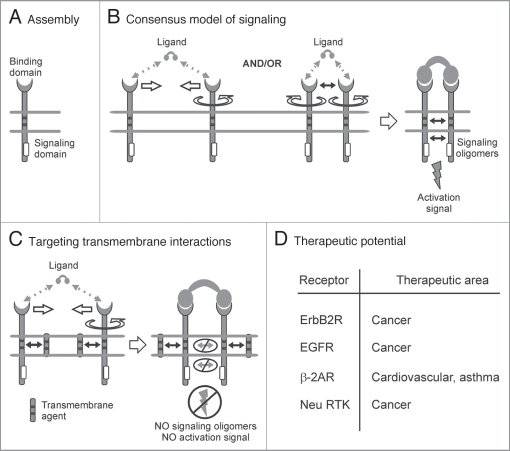
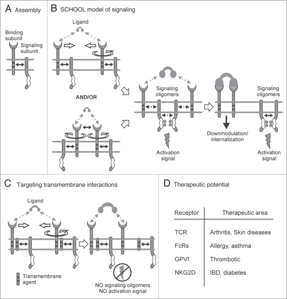
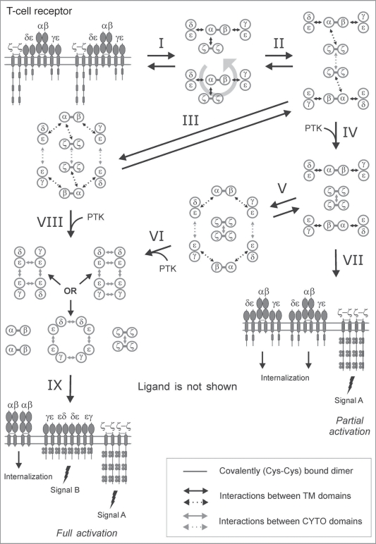
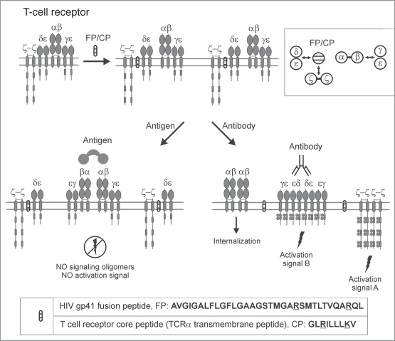
Single-chain receptors and multi-chain immune recognition receptors (SRs and MIRRs, respectively) represent families of structurally related but functionally different surface receptors expressed on different cells. In contrast to SRs, a distinctive and common structural characteristic of MIRR family members is that the extracellular recognition domains and intracellular signaling domains are located on separate subunits. How extracellular ligand binding triggers MIRRs and initiates intracellular signal transduction processes is not clear. A novel model of immune signaling, the Signaling Chain HOmoOLigomerization (SCHOOL) model, suggests that the homooligomerization of receptor intracellular signaling domains represents a necessary and sufficient condition for receptor triggering. In this review, I demonstrate striking similarities between a consensus model of SR signaling and the SCHOOL model of MIRR signaling and show how these models, together with the lessons learned from viral pathogenesis, provide a molecular basis for novel pharmacological approaches targeting inter- and intrareceptor transmembrane interactions as universal therapeutic targets for a diverse variety of immune and other disorders.


Figures

Figure 1
Figure 1
Single-chain receptors. Single activating receptor assembly and signaling. (A) Single-chain receptor (SR) assembly. Extracellular recognition domain and intracellular signaling domain with a signaling sequence (shown by empty rectangles) are located on the same protein chain. (B) Consensus model of SR signaling. The model proposes that receptor homooligomerization in the cytoplasmic milieu plays a central role in triggering SRs. Ligand-induced SR clustering and reorientation (and/or receptor reorientation in preexisting SR clusters) results in SR oligomerization mediated by transmembrane interactions. In these oligomers, receptors are in sufficient proximity and adopt a correct (permissive) relative orientation and geometry to promote homointeractions between cytoplasmic domains. Formation of competent signaling oligomers results in generation of the activation signal (for receptor tyrosine kinases, this means trans-autophosphorylation of Tyr residues in cytoplasmic signaling sequences) and thus triggers downstream signaling pathways. Protein-protein interactions are shown by solid black arrows. Empty arrows illustrate ligand-induced receptor clustering (oligomerization). Circular arrow indicates ligand-induced receptor reorientation. (C) Interreceptor transmembrane interactions. Within the consensus model of SR signaling, specific blockade of interreceptor transmembrane interactions prevents ligand-induced SR oligomerization. Competent signaling oligomers in cytoplasmic milieu are not formed, thus preventing generation of the activation signal. (D) Therapeutic potential of SR transmembrane inhibitors.
Figure 2
Figure 2
Multi-chain immune recognition receptors. Multi-chain activating receptor assembly and signaling. (A) Structural and functional organization of multi-chain immune recognition receptors (MIRRs). Immunoreceptor tyrosine-based activation motif (ITAM) is indicated by empty box. Transmembrane interactions between MIRR ligand-binding and signaling components (shown by solid arrow) play a key role in receptor assembly and integrity on resting cells. (B) The signaling chain homooligomerization (SCHOOL) model, proposing that the homooligomerization of signaling subunits plays a central role in triggering MIRR-mediated signal transduction. Small solid black arrows indicate specific intersubunit hetero- and homointeractions between transmembrane and cytoplasmic domains, respectively. Circular arrow indicates ligand-induced receptor reorientation. Empty arrows illustrate ligand-induced receptor clustering (oligomerization). All interchain interactions in a dimeric intermediate are shown by dotted black arrows reflecting their transition state. Curved lines depict disorder of the cytoplasmic domains of MIRR signaling subunits. Phosphate groups are shown as filled gray circles. (C) Molecular mechanisms underlying proposed intervention by transmembrane-targeted agents. Specific blockade of transmembrane interactions between MIRR recognition and signaling subunits results in “pre-dissociation” of the receptor complex, thus preventing formation of signaling oligomers and inhibiting ligand-dependent immune cell activation. In contrast, stimulation of these “pre-dissociated” MIRRs with cross-linking antibodies to signaling subunit(s) should still lead to receptor triggering and cell activation (not shown). (D) Therapeutic potential of MIRR transmembrane inhibitors.
Figure 3
Figure 3
SCHOOL model of T-cell receptor signaling. A schematic representation of the SCHOOL-based molecular mechanisms of T-cell receptor (TCR) signaling. Immunoreceptor tyrosine-based activation motifs (ITAMs) are shown as gray rectangles. TCR-CD3-ζ components are represented as whole polypeptides and as a simplified axial view. All interchain interactions in intermediate complexes are shown by dotted arrows reflecting their transition state. Circular arrow indicates ligand-induced receptor reorientation. Interaction with multivalent ligand (not shown) clusters the receptors and pushes them to reorientate (I) and bring signaling subunits into a correct relative orientation and in sufficient proximity in the formed receptor oligomer (for illustrative purposes, receptor dimer is shown), thus starting the trans-homointeractions between ζ molecules (II). Then, two alternative pathways can take a place depending on the nature of activating stimuli. First is going through a stage IV resulting in formation of ζ2 dimer (dimer of dimers) and phosphorylation of the ζ ITAM tyrosines, thus triggering downstream signaling events. Then, the signaling ζ oligomers formed subsequently dissociate from the TCR-CD3 complex, resulting in internalization of the remaining engaged TCR-CD3 complexes (VII). This pathway leads to partial (or incomplete) T cell activation. Alternatively, the intermediate complex formed at the stage II can undergo further rearrangements, starting trans-homointeractions between CD3 proteins (III) and resulting in formation of an oligomeric intermediate. Again, the stages I, II and III can be reversible or irreversible depending on interreceptor proximity and relative orientation of the receptors in TCR dimers/oligomers as well as on time duration of the TCR-ligand contact and lifetime of the receptor in TCR dimers/oligomers that generally correlate with the nature of the stimulus and its specificity and affinity/avidity. Next, in the signaling oligomers formed (III), the ITAM tyrosines undergo phosphorylation by PTKs that leads to generation of the activation signal, dissociation of signaling oligomers and internalization of the remaining engaged TCRαβ chains (VIII, XI). This pathway provides at least two different activation signals from the ζ and CD3 signaling oligomers (signals A and B), respectively, and results in full T-cell activation. The distinct signaling through ζ and CD3 oligomers (or through various combinations of signaling chains in CD3 oligomeric structures) might be also responsible for distinct functions such as T-cell proliferation, effector functions, T-cell survival, pathogen clearance, TCR anergy, etc. In addition, the signaling oligomers formed can sequentially interact with the signaling subunits of nonengaged TCRs resulting in formation of higher-order signaling oligomers, thus amplifying and propagating the activation signal (not shown). Also, this leads to the release and subsequent internalization of the remaining nonengaged TCR complexes and/or TCRαβ chains (not shown). Abbreviations: PTK, protein tyrosine kinase. Phosphate groups are shown as filled gray circles.
Figure 4
Figure 4
SCHOOL model of T-cell receptor signaling in the presence of transmembrane peptides. A schematic representation of the SCHOOL-based mechanisms of action of T-cell receptor transmembrane inhibitors such as the T-cell receptor core peptide (CP) and HIV-1 gp41 fusion peptide (FP). Considering the close similarity in patterns of inhibition of T-cell activation and immunosuppressive activity observed for CP and FP, the SCHOOL model reasonably suggests a similar molecular mechanism of action for both peptides. Within the model, these peptides compete with the TCRα chain for binding to the CD3δɛ and ζ signaling subunits, thus disrupting the transmembrane (TM) interactions between these subunits and resulting in disconnection and predissociation of the relevant signaling subunits from the remaining receptor complex (also shown in the inset as a simplified axial view). This prevents formation of signaling oligomers upon multivalent antigen stimulation, thus inhibiting antigen-mediated T-cell activation. In contrast, stimulation of these “predissociated” MIRRs with cross-linking antibodies to signaling subunit(s) should still lead to receptor triggering and cell activation. The model predicts that the same mechanisms of inhibitory action can be applied to TCR TM peptides corresponding to the TM regions of not only the TCRαβ recognition subunits but the corresponding CD3ɛ, CD3δ, CD3γ and ζ signaling subunits as well.
Figure 5
Figure 5
SCHOOL mechanisms of selective modulation of T-cell receptor signaling by different transmembrane peptides. A schematic representation of the SCHOOL-based mechanisms of action of different T-cell receptor transmembrane inhibitors. Within the SCHOOL model, upon antigen stimulation of T cells, T-cell receptor α-chain (TCRα) transmembrane peptide (TMP) prevents formation of all signaling oligomers, including ζ, CD3ɛ, CD3δ and CD3γ. This inhibits T-cell activation in both in vitro and in vivo. In contrast, other TMPs prevent formation of signaling oligomers (and therefore signaling) of selected signaling subunits. This inhibits T-cell activation in vivo whereas inhibition in vitro depends on the evaluation method used. For example, antigen-stimulated induction of cytokine secretion and T-cell proliferation in T cells lacking CD3γ and/or CD3δ cytoplasmic domains, thus explaining the absence of inhibitory effect of the CD3δ and CD3γ TM peptides in the in vitro IL-2 production and T-cell proliferation assays used as markers of T-cell activation. Abbreviations: AD, atopic dermatitis; AIA, adjuvant-induced arthritis; IL-2, interleukin 2.
Figure 6
Figure 6
Similarities in the charge distribution patterns of different immunomodulatory viral sequences. Charge distribution patterns of different immunomodulatory viral sequences. Primary sequence analysis of proven and predicted immunomodulatory sequences of viral fusion protein regions and other domains shows a similarity in charge distribution pattern with two essential positively charged residues spaced apart by 4 (class I) or 8 (class III) amino acids or with three essential positively charged residues spaced apart by 3 amino acids (class II), suggesting a similarity of the SCHOOL-based mechanisms used by diverse viruses in their pathogenesis to modulate the host immune response. Abbreviations: TCR, T-cell receptor; CP, core peptide; HIV, human immunodeficiency virus; gp, glycoprotein; FP, fusion peptide/protein; TMD, transmembrane domain; CKS-17, a synthetic retroviral envelope heptadecapeptide; Fr-MLV, Friend murine leukemia virus; gp, glycoprotein; HHV-6 U24, human herpesvirus 6 U24 protein; HTLV-1, human T lymphotropic virus type 1; HVA, herpesvirus ateles; HVS, herpesvirus saimiri; ITAM, immunoreceptor tyrosine-based activation motif; LASV, Lassa virus; LCMV, lymphocytic choriomeningitis virus; MARV, Marburg virus; MOPV, Mopeia virus; SARS-CoV, severe acute respiratory syndrome coronavirus; SEBOV, Sudan Ebola virus; TACV, Tacaribe virus; Tip, tyrosine kinase interacting protein; Tio, two-in-one protein; TMD, transmembrane domain; ZEBOV, Zaire Ebola virus.

References

    1. Rudd CE. Disabled receptor signaling and new primary immunodeficiency disorders. N Engl J Med. 2006;354:1874–1877. - PubMed
    1. Sigalov AB, editor. Multichain Immune Recognition Receptor Signaling: From Spatiotemporal Organization to Human Disease. New York: Springer-Verlag; 2008. - PubMed
    1. Keegan AD, Paul WE. Multichain immune recognition receptors: similarities in structure and signaling pathways. Immunol Today. 1992;13:63–68. - PubMed
    1. Sigalov AB. Immune cell signaling: a novel mechanistic model reveals new therapeutic targets. Trends Pharmacol Sci. 2006;27:518–524. - PubMed
    1. Heldin CH. Dimerization of cell surface receptors in signal transduction. Cell. 1995;80:213–223. - PubMed

Substances