

Review
doi: 10.4161/cc.9.12.11988.
Epub 2010 Jun 15.
Ubiquitylation and proteasomal degradation of the p21(Cip1), p27(Kip1) and p57(Kip2) CDK inhibitors
Affiliations
- PMID: 20519948
- PMCID: PMC3319752
- DOI: 10.4161/cc.9.12.11988
Item in Clipboard
Review
Ubiquitylation and proteasomal degradation of the p21(Cip1), p27(Kip1) and p57(Kip2) CDK inhibitors
Cell Cycle.
.
Abstract
The expression levels of the p21(Cip1) family CDK inhibitors (CKIs), p21(Cip1), p27(Kip1) and p57(Kip2), play a pivotal role in the precise regulation of cyclin-dependent kinase (CDK) activity, which is instrumental to proper cell cycle progression. The stabilities of p21(Cip1), p27(Kip1) and p57(Kip2) are all tightly and differentially regulated by ubiquitylation and proteasome-mediated degradation during various stages of the cell cycle, either in steady state or in response to extracellular stimuli, which often elicit site-specific phosphorylation of CKIs triggering their degradation.
Figures
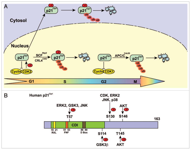
Phosphorylated p21Cip1 is degraded by distinct E3 ligases. (A) E3 ligases involved in p21Cip1 degradation. p21Cip1 is ubiquitylated and degraded in late G1 and S phases by SCFSkp2 and CRL4Cdt2, and in G2 phase by APC/CCdc20 in the nucleus. A portion p21Cip1 is phosphorylated and translocated into the cytosol where it is ubiquitylated and degraded by proteasomes. (B) Schematic structure of p21Cip1 showing the regulatory phosphorylation sites and the cognate protein kinases. CDI: CDK inhibitor domain.
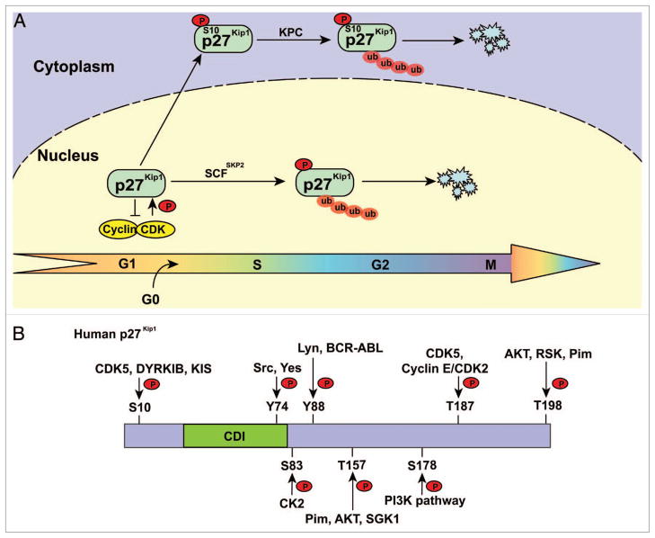
Phosphorylated 27Kip1 is degraded by distinct E3 ligases. (A) E3 ligases involved in p27Kip1 degradation. p27Kip1 is ubiquitylated and degraded in late G1, S and G2 phases by SCFSkp2 in the nucleus. p27Kip1 phosphorylated at S10 is ubiquitylated by the KPC complex when exported to the cytoplasm. (B) Schematic structure of p27Kip1 showing the regulatory phosphorylation sites and the cognate protein kinases. CDI, CDK inhibitor domain.
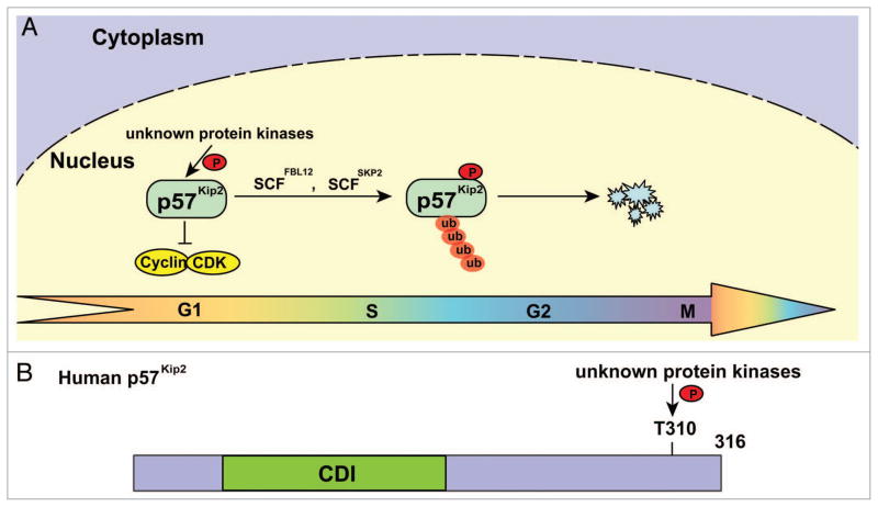
Phosphorylated p57Kip2 is degraded by distinct E3 ligases. (A) E3 ligases involved in p57Kip2 degradation. p57KIP2 phosphorylated at T329 is ubiquitylated and degraded in late G1 and S phases by SCFFBL12 and SCFSkp2. (B) Schematic structure of p57Kip2 showing the single regulatory phosphorylation site. CDI, CDK inhibitor domain.
Similar articles
-
CDK inhibitors, p21(Cip1) and p27(Kip1), participate in cell cycle exit of mammalian cardiomyocytes.Biochem Biophys Res Commun. 2014 Jan 17;443(3):1105-9. doi: 10.1016/j.bbrc.2013.12.109. Epub 2013 Dec 28. Biochem Biophys Res Commun. 2014. PMID: 24380855
-
Expression of p21(Wafl/Cip1), p57(Kip2) and HER2/neu in patients with gallbladder cancer.Anticancer Res. 2007 May-Jun;27(3B):1679-84. Anticancer Res. 2007. PMID: 17595796
-
CDK inhibitors selectively diminish cell cycle controlled activation of the histone H4 gene promoter by p220NPAT and HiNF-P.J Cell Physiol. 2009 May;219(2):438-48. doi: 10.1002/jcp.21687. J Cell Physiol. 2009. PMID: 19170105 Free PMC article.
-
The multiple roles of the cyclin-dependent kinase inhibitory protein p57(KIP2) in cerebral cortical neurogenesis.Dev Neurobiol. 2012 Jun;72(6):821-42. doi: 10.1002/dneu.20999. Dev Neurobiol. 2012. PMID: 22076965 Review.
-
Regulation of p27Kip1 and p57Kip2 Functions by Natural Polyphenols.Biomolecules. 2020 Sep 13;10(9):1316. doi: 10.3390/biom10091316. Biomolecules. 2020. PMID: 32933137 Free PMC article. Review.
Cited by
-
Sensitization of lung cancer cells to cisplatin by β-elemene is mediated through blockade of cell cycle progression: antitumor efficacies of β-elemene and its synthetic analogs.Med Oncol. 2013 Mar;30(1):488. doi: 10.1007/s12032-013-0488-9. Epub 2013 Feb 9. Med Oncol. 2013. PMID: 23397083
-
Deubiquitinase PSMD7 regulates cell fate and is associated with disease progression in breast cancer.Am J Transl Res. 2020 Sep 15;12(9):5433-5448. eCollection 2020. Am J Transl Res. 2020. PMID: 33042429 Free PMC article.
-
Mechanisms of ciliogenesis suppression in dividing cells.Cell Mol Life Sci. 2017 Mar;74(5):881-890. doi: 10.1007/s00018-016-2369-9. Epub 2016 Sep 26. Cell Mol Life Sci. 2017. PMID: 27669693 Free PMC article. Review.
-
Partial hepatectomy in rats results in immediate down-regulation of p27Kip1 in residual liver tissue by transcriptional and post-translational processes.Front Physiol. 2013 Jun 13;4:139. doi: 10.3389/fphys.2013.00139. eCollection 2013. Front Physiol. 2013. PMID: 23781207 Free PMC article.
-
ATM-Mediated translocation of RanBPM regulates DNA damage response by stabilizing p21 in non-small cell lung cancer cells.Cell Oncol (Dordr). 2024 Feb;47(1):245-258. doi: 10.1007/s13402-023-00866-x. Epub 2023 Sep 7. Cell Oncol (Dordr). 2024. PMID: 37676377 Free PMC article.
References
-
- Jackson PK, Eldridge AG, Freed E, Furstenthal L, Hsu JY, Kaiser BK, et al. The lore of the RINGs: substrate recognition and catalysis by ubiquitin ligases. Trends Cell Biol. 2000;10:429–39. - PubMed
-
- Hatakeyama S, Yada M, Matsumoto M, Ishida N, Nakayama KI. U box proteins as a new family of ubiquitin-protein ligases. J Biol Chem. 2001;276:33111–20. - PubMed
-
- Boname JM, Stevenson PG. MHC class I ubiquitination by a viral PHD/LAP finger protein. Immunity. 2001;15:627–36. - PubMed
Publication types
MeSH terms
Substances
Grants and funding
LinkOut - more resources
Full Text Sources
Miscellaneous