. 2010 Jun 3;465(7298):577-83.
doi: 10.1038/nature09141.

HIF-1 antagonizes p53-mediated apoptosis through a secreted neuronal tyrosinase

Affiliations

HIF-1 antagonizes p53-mediated apoptosis through a secreted neuronal tyrosinase

Ataman Sendoel et al. Nature. .

Abstract

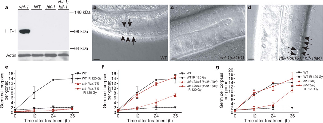
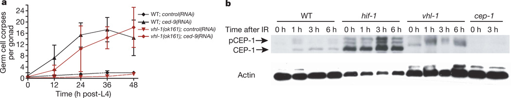
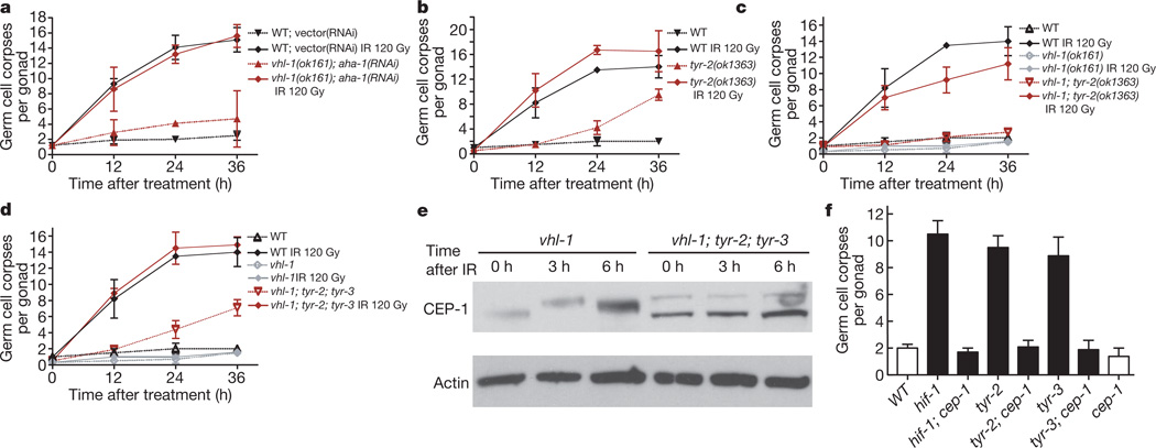
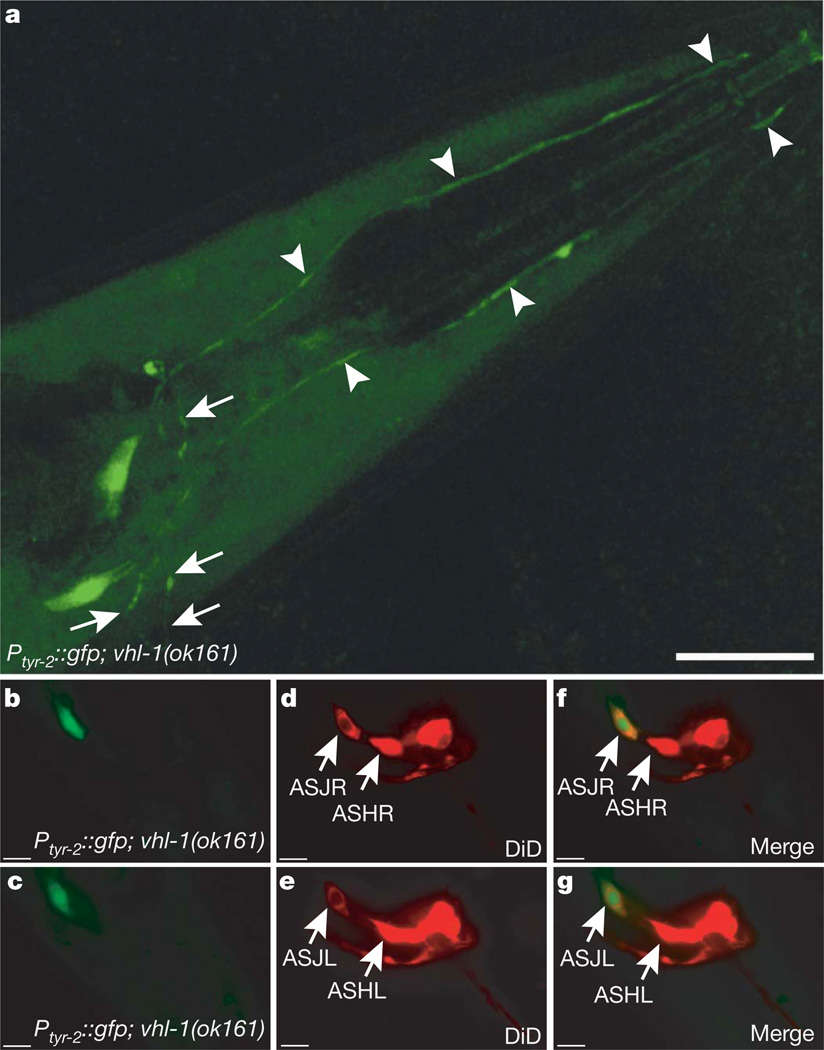
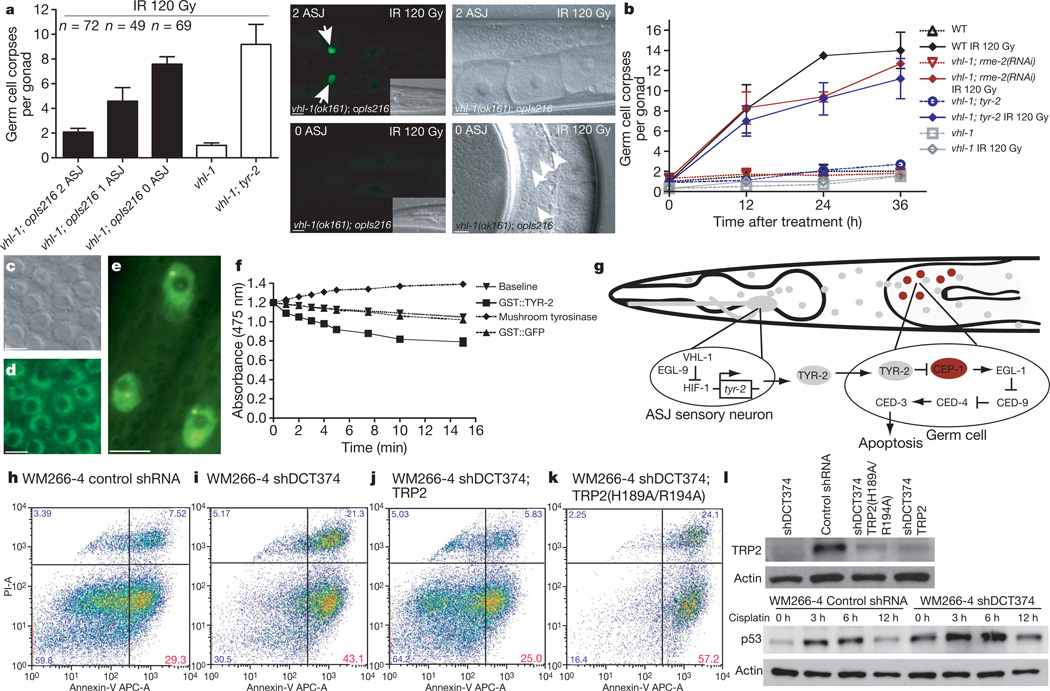
Hypoxia-inducible factor (HIF) is a transcription factor that regulates fundamental cellular processes in response to changes in oxygen concentration. HIFalpha protein levels are increased in most solid tumours and correlate with patient prognosis. The link between HIF and apoptosis, a major determinant of cancer progression and treatment outcome, is poorly understood. Here we show that Caenorhabditis elegans HIF-1 protects against DNA-damage-induced germ cell apoptosis by antagonizing the function of CEP-1, the homologue of the tumour suppressor p53. The antiapoptotic property of HIF-1 is mediated by means of transcriptional upregulation of the tyrosinase family member TYR-2 in the ASJ sensory neurons. TYR-2 is secreted by ASJ sensory neurons to antagonize CEP-1-dependent germline apoptosis. Knock down of the TYR-2 homologue TRP2 (also called DCT) in human melanoma cells similarly increases apoptosis, indicating an evolutionarily conserved function. Our findings identify a novel link between hypoxia and programmed cell death, and provide a paradigm for HIF-1 dictating apoptotic cell fate at a distance.


Figures

Figure 1
Figure 1. HIF-1 antagonizes DNA-damage-induced apoptosis
a, HIF-1 western blot analysis of synchronized young adult hermaphrodites. All alleles used in this study are defined in Methods. bd, Synchronized young adult hermaphrodites were exposed to ionizing radiation (IR) and germline apoptosis was analysed by DIC microscopy 12 h after treatment. Arrows indicate germ cell corpses. Scale bars, 10 µm. eg, Quantification of germline apoptosis. Synchronized young adult hermaphrodites were exposed to IR and germline apoptosis was quantified at the indicated time points. Data shown represent the average of three to six independent experiments ± s.d. (n > 20 animals for each experiment and time point).
Figure 2
Figure 2. HIF-1 impedes DNA-damage-induced apoptosis by inhibiting CEP-1
a, Wild-type or vhl-1(ok161) worms were grown on bacteria containing either an empty control RNAi vector or ced-9(RNAi). Germline apoptosis was quantified at the indicated time points beginning at the fourth larval (L4) stage. Data shown represent the average of three independent experiments ± s.d. (n > 20). b, CEP-1 western blot analysis of synchronized young adult animals. pCEP-1, phosphorylated CEP-1. β-actin was used as loading control.
Figure 3
Figure 3. The antiapoptotic function of HIF-1 is mediated by a concerted action of TYR-2 and TYR-3
a, Wild-type or vhl-1(ok161) worms were grown on bacteria containing either an empty control RNAi vector or the HIF-1β homologue aha-1(RNAi) beginning at the third larval stage (L3). Synchronized young adult animals were irradiated and germline apoptosis was quantified at the indicated time points. Data shown represent the average of three independent experiments ± s.d. (n > 20 per time point and experiment). bd, Time-course analysis of germ cell apoptosis in synchronized control or irradiated young adult animals. Data shown represent the average of three to six independent experiments ± s.d. (n > 20). e, Western blot analysis of CEP-1 in synchronized young adult worms. β-actin was used as loading control. f, Analysis of germ cell apoptosis in unirradiated synchronized animals 48 h after L4 stage. Data shown represent the average of three independent experiments ± s.d. (n > 20).
Figure 4
Figure 4. HIF-1 drives tyr-2 expression in ASJ neurons
a, Transcriptional tyr-2 GFP-reporter opIs216[Ptyr-2::gfp] in a vhl-1(ok161) mutant background reveals HIF-1-dependent expression in two neurons in the head. Arrowheads indicate the dendritic extensions to the anterior end of the worm. Arrows indicate axons that are associated with the nerve ring. Scale bar, 20 µm. bg, DiD staining in opIs216[Ptyr-2::gfp]; vhl-1(ok161) reveals that the GFP signal co-localizes with the most ventro-posterior amphid neurons, the ASJL and ASJR sensory neurons. Scale bars, 10 µm.
Figure 5
Figure 5. TYR-2 is a l-dopachrome tautomerase secreted by the ASJ neurons to inhibit apoptosis
a, Laser microbeam ablation of ASJ neurons. Germline apoptosis of laser-ablated and mock-ablated animals was quantified 24 h after irradiation and the presence of GFP-positive ASJ neurons was determined subsequently. The number of intact ASJ neurons is indicated. Representative head and appendant germ lines are shown on the right; mock-ablated animals in the top row, ASJ-ablated animals are shown in the bottom row. Arrows indicate ASJ neurons; arrowheads apoptotic germ cells. Scale bars, 10 µm. b, Germline endocytosis was inhibited by rme-2(RNAi) and germ cell apoptosis quantified. Data shown represent the average of three independent experiments ± s.d. (n > 20 animals). c, d, DIC microscopy section and TYR-2::GFP nuclear localization in the germ line of Phus-1::tyr-2-SP::gfp::tyr-2(3′ region) animals, which express TYR-2 lacking the signal peptide (amino acids 1–22). Scale bars, 5 µm. e, Nuclear localization of TYR-2::GFP in Ptyr-2::tyr-2-SP::gfp::tyr-2(3′ region) animals. Hypodermal nuclei are depicted. Scale bar, 5 µm. f, l-dopachrome tautomerase activity of GST::TYR-2, GST::GFP and mushroom tyrosinase. g, Model for HIF-1-mediated inhibition of germline apoptosis. hk, Loss of human tyrosinase-related protein 2 (TRP2; also called DCT) results in increased cisplatin-induced apoptosis in WM266-4 metastatic melanoma cells. Melanoma cells were treated with 50 µM cisplatin, stained with Annexin-V/propidium iodide (PI) 24 h after treatment and analysed by flow cytometry. TRP2 knock down (shDCT374) increases Annexin-V positive/PI negative fractions (early apoptotic cells) compared to control shRNA (Luciferase) in WM266-4 melanoma cells (h, i). The shDCT374 (targeting endogenous TRP2 5′ UTR) phenotype is rescued by expression of wild-type TRP2 (MSCV-DCT) (j), but not of the catalytically inactive point mutant TRP2(H189A/R194A) (MSCV-DCT)(k). Data shown are representative of four experiments. APC-A, allophycocyanin conjugate-Annexin-V. l, TRP2 and p53 western blot analysis in WM266-4 melanoma cells.

Comment in

References

    1. Epstein AC, et al. elegans EGL-9 and mammalian homologs define a family of dioxygenases that regulate HIF by prolyl hydroxylation. Cell. 2001;107:43–54. - PubMed
    1. Bruick RK, McKnight SL. A conserved family of prolyl-4-hydroxylases that modify HIF. Science. 2001;294:1337–1340. - PubMed
    1. Ivan M, et al. Biochemical purification and pharmacological inhibition of a mammalian prolyl hydroxylase acting on hypoxia-inducible factor. Proc. Natl Acad. Sci. USA. 2002;99:13459–13464. - PMC - PubMed
    1. Yu F, White SB, Zhao Q, Lee FS. HIF-1α binding to VHL is regulated by stimulus-sensitive proline hydroxylation. Proc. Natl Acad. Sci. USA. 2001;98:9630–9635. - PMC - PubMed
    1. Latif F, et al. Identification of the von Hippel-Lindau disease tumor suppressor gene. Science. 1993;260:1317–1320. - PubMed

Publication types

MeSH terms

Substances