. 2010 May 24;5(5):e10772.
doi: 10.1371/journal.pone.0010772.

Understanding the role of PknJ in Mycobacterium tuberculosis: biochemical characterization and identification of novel substrate pyruvate kinase A

Affiliations

Understanding the role of PknJ in Mycobacterium tuberculosis: biochemical characterization and identification of novel substrate pyruvate kinase A

Gunjan Arora et al. PLoS One. .

Abstract

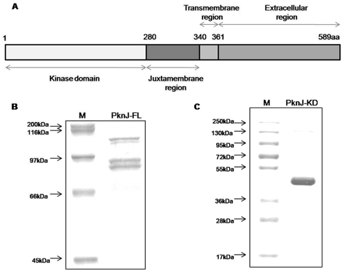
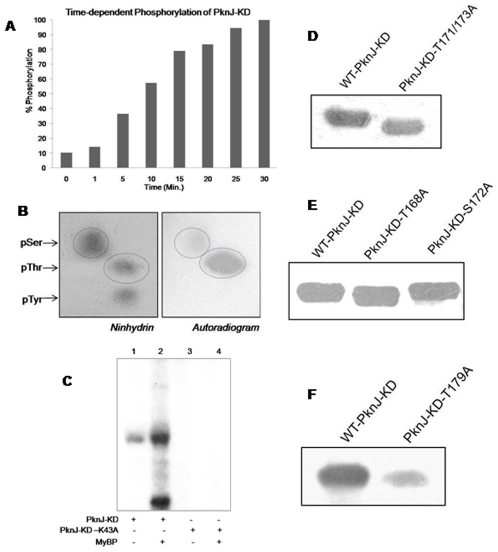
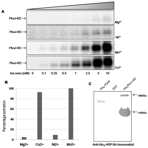
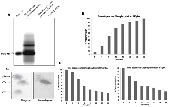
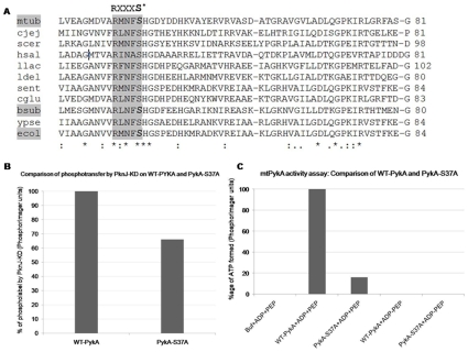
Reversible protein phosphorylation is a prevalent signaling mechanism which modulates cellular metabolism in response to changing environmental conditions. In this study, we focus on previously uncharacterized Mycobacterium tuberculosis Ser/Thr protein kinase (STPK) PknJ, a putative transmembrane protein. PknJ is shown to possess autophosphorylation activity and is also found to be capable of carrying out phosphorylation on the artificial substrate myelin basic protein (MyBP). Previous studies have shown that the autophosphorylation activity of M. tuberculosis STPKs is dependent on the conserved residues in the activation loop. However, our results show that apart from the conventional conserved residues, additional residues in the activation loop may also play a crucial role in kinase activation. Further characterization of PknJ reveals that the kinase utilizes unusual ions (Ni(2+), Co(2+)) as cofactors, thus hinting at a novel mechanism for PknJ activation. Additionally, as shown for other STPKs, we observe that PknJ possesses the capability to dimerize. In order to elucidate the signal transduction cascade emanating from PknJ, the M. tuberculosis membrane-associated protein fraction is treated with the active kinase and glycolytic enzyme Pyruvate kinase A (mtPykA) is identified as one of the potential substrates of PknJ. The phospholabel is found to be localized on serine and threonine residue(s), with Ser(37) identified as one of the sites of phosphorylation. Since Pyk is known to catalyze the last step of glycolysis, our study shows that the fundamental pathways such as glycolysis can also be governed by STPK-mediated signaling.


Conflict of interest statement

Competing Interests: The authors have declared that no competing interests exist.

Figures

Figure 1
Figure 1. Domain architecture and protein purification of full length and kinase domain of M. tuberculosis PknJ (Rv2088).
(A) A schematic representation of PknJ domain organization. The domains were predicted using TMHMM (http://www.cbs.dtu.dk/services/TMHMM/), HMMTOP (http://www.enzim.hu/hmmtop/) and DAS (http://www.sbc.su.se/~miklos/DAS/). (B) Full length PknJ was purified as GST-tagged protein of ∼93 kDa which migrates as duplet. Purified GST-PknJ-FL was run on 8% SDS-PAGE and stained with Coomassie Brilliant Blue. (C) Cytosolic kinase domain of PknJ containing 1–320 amino acids was purified as His6-tagged protein of 39 kDa which migrates at a higher size of ∼43 kDa. Purified His6-PknJ-KD was run on 12% SDS-PAGE and stained with Coomassie brilliant blue.
Figure 2
Figure 2. In vitro kinase assay of PknJ, Phosphoamino acid analysis and Immunoblotting of PknJ-KD and activation loop mutants.
(A) Graph showing time-dependent autophosphorylation of PknJ-KD. After in vitro kinase assay with [γ-32P]ATP, gel was analyzed by PhosphorImaging and counts were calculated with MultiGauge (FujiFilm). Counts obtained after 30 min of reaction were taken as 100% signal and relative phosphorylation was estimated. Experiment was performed twice and the results indicate average of the two. (B) Analysis of phosphoamino acid content of autophosphorylated PknJ-KD. Amino acid standards, phosphoserine (pSer), phosphothreonine (pThr) and phosphotyrosine (pTyr) were added in the radiolabeled sample and visualized by ninhydrin staining (left panel) prior to autoradiography (right panel). The labeled pThr, pSer and their corresponding standards are encircled. (C) In vitro autophosphorylation of PknJ-KD (1 µg) and phosphotransfer on 5 µg Myelin basic protein (MyBP). Autophosphorylation deficient mutant PknJ-KD-K43A was used as negative control for auto- and transphosphorylation. The reactions were run on 12% SDS-PAGE and gel was autoradiographed after drying. As shown, transphosphorylation on MyBP was visible only in the presence of PknJ-KD, lane2. (D, E, F) 2 µg of in vitro phosphorylated native kinase along with its activation loop mutants (as indicated) were resolved on 10% SDS-PAGE, transferred onto nitrocellulose membrane and probed with anti-phosphothreonine antibody as described in experimental procedures. Autoradiograms are shown.
Figure 3
Figure 3. Regulation of PknJ-KD by metals-ions and dimer formation.
(A) 1 µg PknJ-KD was incubated with increasing concentrations (0–10 mM) of different metal ions, individually in in vitro kinase assays. (B) Comparative analysis of PknJ-KD activation by MgCl2, MnCl2, CoCl2 and NiCl2 (10 mM each). Quantification of PhosphorImager units was done using ImageGauge software (Fujifilm) and converted to percentage activation. (C) To show PknJ-KD dimerization by immunoblotting, excess of PknJ-KD was resolved on 10% SDS-PAGE, transferred onto nitrocellulose membrane and probed with anti-His5HRP-conjugated antibody. His6-PykA and GST were taken as positive and negative controls, respectively.
Figure 4
Figure 4. In vitro phosphorylation of membrane-associated protein fraction and mtPykA by PknJ-KD.
(A) 20 µg purified fraction of membrane-associated proteins was incubated with 2 µg of PknJ-KD or PknJ-KD-K43A, in an in vitro kinase assay. Samples were separated by 10% SDS-PAGE and autoradiographed on PhosphorImager. (B) Graph showing time-dependent phosphorylation of mtPykA by PknJ-KD. 1 µg of PknJ-KD was incubated with 2 µg of mtPykA with increasing time-points (0–30minutes) in in vitro kinase assay. Phosphorylation was evaluated as before. Experiment was performed twice and the results indicate average of the two. (C) Phosphoamino acid content of mtPykA phosphorylated by PknJ-KD was assessed as discussed earlier. (D) Time-dependent dephosphorylation of in vitro phosphorylated PknJ-KD (left panel) and mtPykA (right panel) by Mstpcat. Extent of dephosphorylation was measured by adding 500 ng of purified Mstpcat to a reaction mixture containing phosphorylated PknJ-KD and mtPykA for increasing time-points. Dephosphorylation was evaluated as discussed before with signal intensity at 0 minute taken as maximum. Experiment was performed twice and the results indicate average of the two.
Figure 5
Figure 5. Importance of mtPykA Ser37 as a target site of PknJ-KD and as a critical residue for mtPykA activity.
(A) Multiple sequence alignment of Pyruvate kinase from various microbial species using t-coffee server (http://www.ebi.ac.uk/Tools/t-coffee/index.html). The conserved RXXXS* motif is highlighted with S* being the phosphorylated residue in M. tuberculosis, E. coli and B. subtilis is shaded grey. Pyk sequences were taken from following species: Mycobacterium tuberculosis (mtub.), Campylobacter jejuni (cjej.), Saccharomyces cerevisiae (scer.), Halobacterium salinarum (hsal.), Lactococcus lactis (llac.), Lactobacillus delbrueckii (ldel.), Salmonella enterica (sent.), Corynebacterium glutamicum (cglu.), bacillus subtilis (bsub.), Yersinia pseudotuberculosis (ypse.) and Escherichia coli (ecol.). (B) Loss of phospholabel on PykA-S37A with respect to WT-PykA. Similar concentrations of WT-PykA and PykA-S37A (2 µg) were incubated with 1 µg PknJ-KD in the presence of [γ-32P]ATP. Image was analyzed by ImageGauge as discussed before. (C) Comparison of WT-PykA and PykA-S37A in terms of ATP generation. Activity assays were performed with [α-32P]ADP and analyzed by ImageGauge.

Similar articles

Cited by

References

    1. Cole ST, Brosch R, Parkhill J, Garnier T, Churcher C, et al. Deciphering the biology of Mycobacterium tuberculosis from the complete genome sequence. Nature. 1998;393:537–544. - PubMed
    1. Bach H, Wong D, Av-Gay Y. Mycobacterium tuberculosis PtkA is a novel protein tyrosine kinase whose substrate is PtpA. Biochem J. 2009;420:155–160. - PubMed
    1. Vohra R, Gupta M, Chaturvedi R, Singh Y. Attack on the scourge of Tuberculosis: Patented Drug Targets. Recent Pat Antiinfect Drug Discov. 2006;1:95–106. - PubMed
    1. Chaba R, Raje M, Chakraborti PK. Evidence that a eukaryotic-type serine/threonine protein kinase from Mycobacterium tuberculosis regulates morphological changes associated with cell division. Eur J Biochem. 2002;269:1078–1085. - PubMed
    1. Av-Gay Y, Jamil S, Drews SJ. Expression and characterization of the Mycobacterium tuberculosis serine/threonine protein kinase PknB. Infect Immun. 1999;67:5676–5682. - PMC - PubMed

Publication types

LinkOut - more resources