. 2010 May 24;5(5):e10787.
doi: 10.1371/journal.pone.0010787.

Active chromatin marks are retained on X chromosomes lacking gene or repeat silencing despite XIST/Xist expression in somatic cell hybrids

Affiliations

Active chromatin marks are retained on X chromosomes lacking gene or repeat silencing despite XIST/Xist expression in somatic cell hybrids

Nancy P Thorogood et al. PLoS One. .

Abstract

Background: X-chromosome inactivation occurs early in mammalian development and results in the inactive X chromosome acquiring numerous hallmarks of heterochromatin. While XIST is a key player in the inactivation process, the method of action of this ncRNA is yet to be determined.

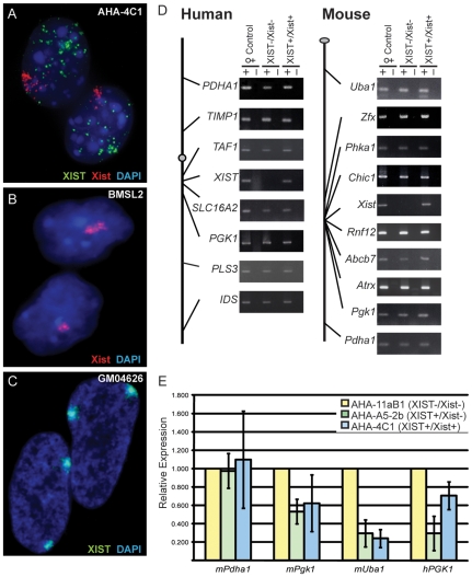
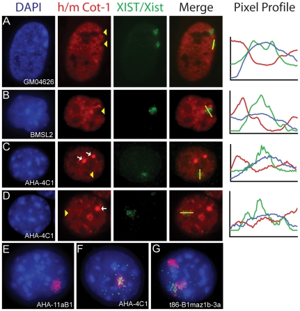
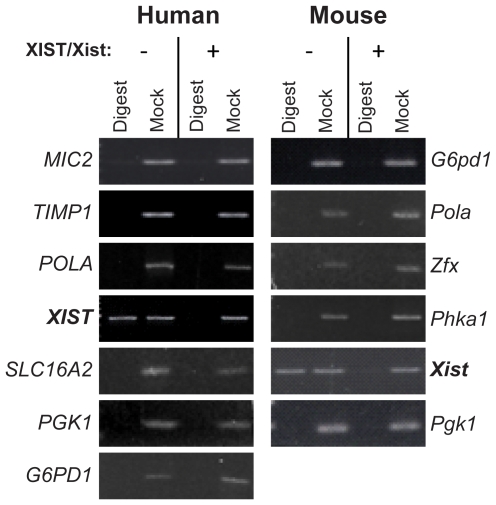
Methodology/principal findings: To assess which features of heterochromatin may be directly recruited by the expression and localization of the XIST RNA we have analyzed a mouse/human somatic cell hybrid in which expression of human and mouse XIST/Xist has been induced from the active X by demethylation. Such hybrids had previously been demonstrated to disconnect XIST/Xist expression from gene silencing and we confirm maintenance of X-linked gene expression, even close to the Xist locus, despite the localized expression of mouse Xist.

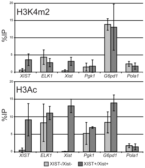
Conclusions/significance: Loss of the active chromatin marks H3 acetylation and H3 lysine 4 methylation was not observed upon XIST/Xist expression, nor was there a gain of DNA methylation; thus these marks of facultative heterochromatin are not solely dependent upon Xist expression. Cot-1 holes, regions of depleted RNA hybridization with a Cot-1 probe, were observed upon Xist expression; however, these were at reduced frequency and intensity in these somatic cells. Domains of human Cot-1 transcription were observed corresponding to the human chromosomes in the somatic cell hybrids. The Cot-1 domain of the X was not reduced with the expression of XIST, which fails to localize to the human X chromosome in a mouse somatic cell background. The human inactive X in a mouse/human hybrid cell also shows delocalized XIST expression and an ongoing Cot-1 domain, despite X-linked gene silencing. These results are consistent with recent reports separating Cot-1 silencing from genic silencing, but also demonstrate repetitive element expression from an otherwise silent X chromosome in these hybrid cells.


Conflict of interest statement

Competing Interests: The authors have declared that no competing interests exist.

Figures

Figure 1
Figure 1. X-linked gene expression in somatic cell hybrids before and after expression of XIST/Xist.
(A–C) Shows the XIST/Xist expression in cell nuclei using RNA FISH probes for the human XIST RNA (green) and the mouse Xist RNA (red). (A) The XIST+/Xist+ (AHA-4C1) somatic cell hybrid; (B) The female mouse fibroblast control cell line, BMSL2; (C) The human female fibroblast line GM04626 (karyotype: 47,XXX). (D) Schematic diagram of both the human and mouse X chromosomes showing the location of the genes analyzed to the left of gel images of RT-PCR products of cDNA from the control female (GM7350 (human) and BMSL2 (mouse)); the hybrid not expressing AHA-11aB1 (XIST/Xist); and the derivative hybrid that expresses AHA-4C1 (XIST+/Xist+). (E) Quantitative RT-PCR of 4 X-linked genes. An intermediate hybrid AHA-A5-2b (XIST+/Xist) was analyzed for expression along with the hybrid AHA-4C1 (XIST+/Xist+). The gene expression was normalized to Actin and expressed as a fold-change relative to the parent hybrid AHA-11aB1 (XIST/Xist). The error bars represent the standard deviation of three separate RNA isolations.
Figure 2
Figure 2. Analysis of Cot-1 hybridization in somatic cell hybrids expressing Xist/XIST by dual RNA FISH hybridization.
Cot-1 RNA (mouse (m) or human (h)) signal is red, Xist/XIST RNA signal is green and DAPI is blue. (A–D) Lines were drawn in ImageJ (NIH) and pixel intensities of each signal was plotted and shown to the right of the images. Yellow arrow heads indicate where the Xist signal corresponds to reductions in the Cot-1 signal. (A) Human female cell line, GM04626, with two inactive X chromosomes and two hCot-1 holes corresponding to the two XIST RNA signals. (B) Mouse female cell line, BMSL2, showing one mCot-1 hole corresponding to the mouse Xist signal, this pattern was observed in 59% of cells (n = 100). (C) XIST+/Xist+ somatic cell hybrid, AHA-4C1, showing a mCot-1 hole corresponding to the mouse Xist signal, this pattern was observed in 7% of cells (n = 100). (D) AHA-4C1 without a mCot-1 hole. In panels C and D bright foci of mCot-1 staining are observed (white arrows). (E) hCot-1 RNA expression (red) in the XIST/Xist hybrid cell, AHA-11aB1, containing only one human chromosome (the X chromosome). (F) hCot-1 RNA expression (red) in the XIST+/Xist+ hybrid cell, AHA-4C1, containing only one human chromosome (the X chromosome), and XIST RNA (green) drifting from the hCot-1 domain. (G) An Xi-hybrid cell line, t86-B1maz1b-3a, containing two human chromosome (X+15). Both human chromosomes form an hCot-1 transcriptional domain, one with the XIST signal drifting away.
Figure 3
Figure 3. DNA methylation of X-linked gene promoters before and after expression of XIST/Xist.
Genomic DNA was cut with a methylation-sensitive restriction enzyme prior to amplification by PCR with primers flanking the enzyme cut sites (lanes labeled Digest). Mock digested samples were used as a positive control (Mock). If a band occurs in the digest lane, it means that the site/sites within the amplicon are methylated (e.g. XIST/Xist). If no band was observed after PCR then we conclude that there was no intact template to amplify as the internal restriction enzyme sites were not methylated.
Figure 4
Figure 4. Active histone modifications at the promoters of X-linked genes after expression of XIST/Xist.
H3K4m2 (upper) and H3Ac (lower) marks were assessed by ChIP followed by quantitative PCR for the two human and four mouse genes listed. The %IP is relative to input and error bars indicate error between 2–3 replicate immunoprecipitations.

Similar articles

Cited by

References

    1. Heard E, Disteche CM. Dosage compensation in mammals: fine-tuning the expression of the X chromosome. Genes Dev. 2006;20:1848–1867. - PubMed
    1. Chaumeil J, Le Baccon P, Wutz A, Heard E. A novel role for Xist RNA in the formation of a repressive nuclear compartment into which genes are recruited when silenced. Genes Dev. 2006;20:2223–2237. - PMC - PubMed
    1. Clemson CM, Hall LL, Byron M, McNeil J, Lawrence JB. The X chromosome is organized into a gene-rich outer rim and an internal core containing silenced nongenic sequences. Proc Natl Acad Sci USA. 2006;103:7688–7693. - PMC - PubMed
    1. Huynh KD, Lee JT. Inheritance of a pre-inactivated paternal X chromosome in early mouse embryos. Nature. 2003;426:857–862. - PubMed
    1. Wutz A, Jaenisch R. A shift from reversible to irreversible X inactivation is triggered during ES cell differentiation. Mol Cell. 2000;5:695–705. - PubMed

Publication types