. 2010 May 24;5(5):e10800.
doi: 10.1371/journal.pone.0010800.

B7-H1-deficiency enhances the potential of tolerogenic dendritic cells by activating CD1d-restricted type II NKT cells

Affiliations

B7-H1-deficiency enhances the potential of tolerogenic dendritic cells by activating CD1d-restricted type II NKT cells

Carolin Brandl et al. PLoS One. .

Abstract

Background: Dendritic cells (DC) can act tolerogenic at a semi-mature stage by induction of protective CD4(+) T cell and NKT cell responses.

Methodology/principal findings: Here we studied the role of the co-inhibitory molecule B7-H1 (PD-L1, CD274) on semi-mature DC that were generated from bone marrow (BM) cells of B7-H1(-/-) mice and applied to the model of Experimental Autoimmune Encephalomyelitis (EAE). Injections of B7-H1-deficient DC showed increased EAE protection as compared to wild type (WT)-DC. Injections of B7-H1(-/-) TNF-DC induced higher release of peptide-specific IL-10 and IL-13 after restimulation in vitro together with elevated serum cytokines IL-4 and IL-13 produced by NKT cells, and reduced IL-17 and IFN-gamma production in the CNS. Experiments in CD1d(-/-) and Jalpha281(-/-) mice as well as with type I and II NKT cell lines indicated that only type II NKT cells but not type I NKT cells (invariant NKT cells) could be stimulated by an endogenous CD1d-ligand on DC and were responsible for the increased serum cytokine production in the absence of B7-H1.

Conclusions/significance: Together, our data indicate that BM-DC express an endogenous CD1d ligand and B7-H1 to ihibit type II but not type I NKT cells. In the absence of B7-H1 on these DC their tolerogenic potential to stimulate tolerogenic CD4(+) and NKT cell responses is enhanced.


Conflict of interest statement

Competing Interests: The authors have declared that no competing interests exist.

Figures

Figure 1
Figure 1. WT and B7-H1−/− DC show equal expression of surface markers and have the same potential to stimulate CD4+ T cells.
A) BM-derived DC from WT and B7-H1−/− mice were matured with TNF over night, stained for different surface markers and analyzed by flow cytometry. The shaded histograms show the unstimulated control and the black lines show TNF-matured DC. Results are representative for 3 independent experiments. B) TNF-stimulated and MOG35–55-loaded DC from WT and B7-H1−/− mice were co-cultured with CFSE-stained CD4+ T cells with MOG35–55 transgenic TCR. Proliferation of the T cells was analysed by the dilution of the CFSE-staining. The dottet line shows the control without DC, the shaded histogram shows the co-culture with 1×10⁴ DC and the filled line shows the co-culture with 3×10⁴ DC. Results are representative for 3 indepentent experiments.
Figure 2
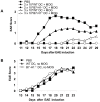
Figure 2. B7-H1−/− DC have a higher tolerogenic potential than WT DC.
A) 2×10⁶ or 3×10⁶ MOG35–55 loaded and TNF-matured WT or B7-H1−/− DC were injected i.v. into WT mice 7, 5 and 3 days before EAE induction. Control mice were injected with PBS. On day 0, EAE was induced and disease course was monitored daily. Results represent the average disease score of 4 mice per group and are representative for 4 independent experiments. B) 2,5×10⁶ TNF-matured, but not MOG35–55 loaded WT or B7-H1−/− DC were injected i.v. into WT mice 7, 5 and 3 days before EAE induction. Results represent the average disease score of 4 mice per group and are representative for 2 independent experiments.
Figure 3
Figure 3. Injection of B7-H1−/− DC results in reduced frequency of neurantigen-specific IFN-γ and IL-17 secreting cells in the CNS.
MOG35–55 loaded and TNF-matured WT or B7-H1−/− DC (2,5×10⁶) were injected i.v. into WT mice 7, 5 and 3 days before EAE induction. Control mice were injected with PBS. After 15 days CNS mononuclear cells were harvested and CNS T cell infitration and cytokine production were investigated using flow cytometry and ELISPOT assay. A) Dot blots are gated for live lymphocytes and inserted numbers represent the percentage of CD4+ and CD8+ CNS infiltrating T cells of 3–5 pooled mice per group of one experiment. Results are representative for 2 independent experiments. B) ELISPOT analysis of IFN-γ (left) or IL-17 (right) production by infiltrating CNS cells after MOG35–55 peptide restimulation. Results are representative for 2 independent experiments with 3–5 pooled mice per group. * p<0.05, ** p<0.01, n.s. not significant.
Figure 4
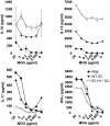
Figure 4. B7-H1−/− DC induce more protective cytokines than WT DC.
MOG35–55 loaded and TNF-matured WT or B7-H1−/− DC (2,5×10⁶) were injected i.v. into WT mice 7, 5 and 3 days before EAE induction. Control mice were injected with PBS. 10 days after EAE induction spleen cells were isolated and restimulated with different concentrations of MOG35–55. After 4 days of culture supernatants were tested by ELISA for IL-10, IL-13, IL-17 and IFN-γ. Results show the mean of 4 mice per group and are representative for 2 independent experiments with 4 mice per group.
Figure 5
Figure 5. Strong increase of protective serum cytokines after injection of B7-H1−/− DC injection is mainly produced by type II NKT cells.
A) Splenocytes from WT and Jα281−/− mice were stained for CD3 and NK1.1 and investigated by flow cytometry. Double positive cells (NKT cells) were analyzed for PD-1 expression. B) MOG35–55 loaded and TNF-matured WT or B7-H1−/− DC (2,5×10⁶) were injected i.v. into WT, CD1d−/− or Jα281−/− mice three times (on day −4, −2 and 0). Similarly, control mice were injected with PBS. Blood was collected 2 hours after the last DC injection and serum cytokines were measured by ELISA for IFN-γ, IL-17, IL-4 and IL-13. The results represent the average of 3 mice per group and are representative for 4 independent experiments for the WT mice and for 2 independent experiments for CD1d−/− and Jα281−/− mice. * p<0.05, ** p<0.01, *** p<0.001, n.s. not significant.
Figure 6
Figure 6. NKT cell hybridoma cells express PD-1 but only the type II NKT cells are stimulated by DC and are negatively regulated by B7-H1.
A) NKT cell hybridoma cells s were stained for PD-1 and analyzed by FACS. The shaded histogram shows the isotype control staining and the black line shows PD-1 staining. B) DC from WT and B7-H1−/− mice were co-cultured with the indicated NKT hybridoma cells in the presence or absence of αGC (10 ng/ml). After 24 hours, the supernatants were tested for IL-2 content by ELISA. Results show 1 out of 4 representative and independent experiments. ** p<0.01.

Similar articles

Cited by

References

    1. Lutz MB, Schuler G. Immature, semi-mature and fully mature dendritic cells: which signals induce tolerance or immunity? Trends Immunol. 2002;23:445–449. - PubMed
    1. Wiethe C, Schiemann M, Busch D, Haeberle L, Kopf M, et al. Interdependency of MHC class II/self-peptide and CD1d/self-glycolipid presentation by TNF-matured dendritic cells for protection from autoimmunity. J Immunol. 2007;178:4908–4916. - PubMed
    1. Menges M, Rossner S, Voigtlander C, Schindler H, Kukutsch NA, et al. Repetitive injections of dendritic cells matured with tumor necrosis factor alpha induce antigen-specific protection of mice from autoimmunity. J Exp Med. 2002;195:15–21. - PMC - PubMed
    1. Pardoll DM. Spinning molecular immunology into successful immunotherapy. Nat Rev Immunol. 2002;2:227–238. - PubMed
    1. Carreno BM, Carter LL, Collins M. Therapeutic opportunities in the B7/CD28 family of ligands and receptors. Curr Opin Pharmacol. 2005;5:424–430. - PubMed

Publication types

MeSH terms