. 2010 May 26;5(5):e10786.
doi: 10.1371/journal.pone.0010786.

Responsiveness to PI3K and MEK inhibitors in breast cancer. Use of a 3D culture system to study pathways related to hormone independence in mice

Affiliations

Responsiveness to PI3K and MEK inhibitors in breast cancer. Use of a 3D culture system to study pathways related to hormone independence in mice

Maria Laura Polo et al. PLoS One. .

Abstract

Background: A significant proportion of breast cancer patients face failure of endocrine therapy due to the acquisition of endocrine resistance. We have explored mechanisms involved in such disease progression by using a mouse breast cancer model that is induced by medroxyprogesterone acetate (MPA). These tumors transit through different stages of hormone sensitivity. However, when cells from tumor variants were seeded on plastic, all were stimulated by progestins and inhibited by antiprogestins such as RU486. Furthermore, cells from a RU486-resistant tumor variant recovered antiprogestin sensitivity.

Hypothesis: A three-dimensional (3D) culture system, by maintaining differential cellular organization that is typical of each tumor variant, may allow for the maintenance of particular hormone responses and thus be appropriate for the study of the effects of specific inhibitors of signaling pathways associated with disease progression.

Method: We compared the behavior of tumors growing in vivo and cancer cells ex vivo (in 3D Matrigel). In this system, we evaluated the effects of kinase inhibitors and hormone antagonists on tumor growth.

Principal findings: LY294002, a PI3K/AKT pathway inhibitor, decreased both tumor growth in vivo and cell survival in Matrigel in MPA-independent tumors with higher AKT activity. Induction of cell death by anti-hormones such as ICI182780 and ZK230211 was more effective in MPA-dependent tumors with lower AKT activity. Inhibition of MEK with PD98059 did not affect tumor growth in any tested variant. Finally, while Matrigel reproduced differential responsiveness of MPA-dependent and -independent breast cancer cells, it was not sufficient to preserve antiprogestin resistance of RU486-resistant tumors.

Conclusion: We demonstrated that the PI3K/AKT pathway is relevant for MPA-independent tumor growth. Three-dimensional cultures were useful to test the effects of kinase inhibitors on breast cancer growth and highlight the need for in vivo models to validate experimental tools used for selective therapeutic targeting.


Conflict of interest statement

Competing Interests: The authors have declared that no competing interests exist.

Figures

Figure 1
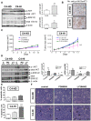
Figure 1. The PI3K/AKT pathway regulates growth in C4-HI but not in C4-HD tumors.
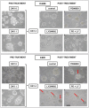
C4-HD tumors were transplanted into BALB/c females carrying a MPA depot while C4-HI tumors were transplanted in the absence of MPA. When tumors reached a 150 mm2 of size, they were removed and processed as described in Materials and Methods for western blot or immunohistochemistry. A. Left. Western blots of protein extracts from C4-HD and C4-HI tumors using specific antibodies against phosphorylated Ser473 AKT (p-AKT), total AKT, phosphorylated ERK1/2 (p-ERK1/2), total ERK1/2 and β-actin (loading control). Three representative samples of a total of six of each tumor type are shown. Right. Quantification of AKT activation (p-AKT relative to total AKT levels) and ERK activation (p-ERK relative to total ERK levels). n = 6 for each tumor type; *: p<0.05 C4-HI vs. C4-HD. B. Immunohistochemistry of slices of paraffin embedded C4-HD or C4-HI tumors using antibodies against phosphorylated Ser473 AKT, showing a higher number of p-AKT-positive cells in C4-HI than in C4-HD tumors. Brown corresponds to the antibody signal and blue marks nuclei stained with hematoxylin. Scale bar: 60 µm. C. 3.6 mg/kg PD98059 (MEK inhibitor), 4 mg/kg LY294002 (PI3K inhibitor) or 100 µl of saline solution (c, control) were administrated i.p. every other day for 12 days to animals carrying C4-HD or C4-HI tumors. Length and width of mammary tumors (mm2) were measured every 2 days. Treatments with the inhibitors started once the tumors reached a size of approximately 30 mm2. Treatment with LY294002 causes C4-HI tumors to have a decreased growth rate. n = 6 for each group; **: p<0.01 vs. control. D. Left. At the end of 12 days of treatment, tumors were removed to evaluate by western blot the inhibitory effect of PD98059 and LY294002 on p-ERK1/2 and p-AKT, respectively. Two representative samples of a total of six are shown in the gel (left). Right. Quantification of AKT and ERK activation as in A. n = 6 for each group; a,b: p<0.01 C4-HI vs. C4-HD; *:p<0.05 vs. control. E. Quantification of the percentage of apoptosis at the end of treatment. Data corresponds to the mean±SEM percentage of apoptotic cells in ten high-power fields corresponding to six independent experiments. Apoptosis is higher in C4-HI tumors treated with LY294002. **: p<0.01 vs. control. F. C4-HI tumors are more differentiated than C4-HD tumors. After 12 days of treatment with LY294002, there is an increase in the number of ductal-like structures only in the C4-HI tumors, as indicated by the black arrows. Scale bar: 30 µm.
Figure 2
Figure 2. Differential sensitivity to PI3K/AKT pathway regulation is lost in isolated cancer cells.
A. Phase contrast microscopy of primary cultures isolated from C4-HD and C4-HI tumors showing flattened monolayers on tissue culture plastic. Scale bar: 30 µm. B. Top. Western blots showing p-AKT, total AKT, p-ERK1/2, ERK1/2 and β-actin in protein extracts from primary cells cultured for 96 hrs. Three representative samples of a total of six are shown in the gel. Bottom. Quantification of p-AKT/total AKT and p-ERK/total ERK levels, n = 6 for each cell type. C. Proliferation assay (3H-thymidine incorporation) in primary C4-HD (left) or C4-HI (right) cells incubated during the last 48 hrs with 0.01 µM MPA and 5, 10 and 20 µM PD98059 or LY294002. For the combined treatment, the inhibitor was used at a dose of 10 µM. 3H-thymidine was added in the last 18 hrs before harvesting the cells. MPA has a stimulatory effect while PD98059 and LY294002 have an inhibitory effect on MPA-treated and untreated cells. n = 8 for each treatment. ***:p<0.001; **:p<0.01; *: p<0.05 vs. control (untreated cells); different letters indicate significant differences between treatments in the presence of MPA with p<0.01. A representative experiment of a total of three is shown here. D. Left. Western blots showing that phosphorylation levels of AKT and ERK1/2 decrease in primary C4-HI cultures incubated for 48 hrs with 10 µM of specific inhibitors. β-actin levels in the blot serve as a loading control. Two representative samples of a total of six are shown (left). Right. Quantification of p-AKT/total AKT and p-ERK/total ERK levels. n = 6 for each group; *p<0.05; **p<0.01 vs. control.
Figure 3
Figure 3. Differential activation of the PI3K/AKT pathway in tumor cells is cell context dependent.
Primary cancer cells in 3D cultures “on top” of Matrigel reproduced the architecture typical of the tumors, as well as the differential pattern of protein kinase activation between cells types. A. C4-HD and C4-HI primary cells isolated from the tumor and placed on tissue culture plastic for 48 hrs show diffuse and irregular deposition of integrin α6 as assayed by immunofluorescence under confocal microscopy. B. Phase contrast microscopy (top) of primary cells placed on top of a thin layer of Matrigel. After 48 hrs, the cells show differences in organization among the three cell types. Confocal images from immunofluorescence studies (bottom) reveal that C4-HI cells (right) form organized clusters with an appropriate continuous basal localization of integrin α6, resembling normal mammary organoids in culture. C4-HI cells exhibit apical localization of MUC-1 on the surface of the cell membrane, lateral localization of tight junction protein ZO-1 and even the presence of a central lumen in some of the clusters. C4-HD cells (left) remain as disorganized clusters, with no clear distribution of any polarity marker tested and no central lumen. Polarity markers were stained green with specific antibodies for immunofluorescence; nuclei were stained red with propidium iodide. Representative clusters of each condition are shown here. Scale bar: 30 µm. C. Top. Western blot of protein extracts obtained from primary cultures grown on Matrigel showing that differences on p-AKT and p-ERK1/2 levels between C4-HD and C4-HI tumors is preserved when isolated cells are placed on 3D culture system. β-actin levels in the blot serve as a loading control. Three representative samples of a total of six are shown. Bottom. Quantification of p-AKT/total AKT and p-ERK/total ERK levels. n = 6 for each cell type; **p<0.01 C4-HI vs. C4-HD.
Figure 4
Figure 4. Higher sensitivity of C4-HI tumors to inhibition of PI3K/AKT pathway is restored in 3D Matrigel.
The 3D Matrigel culture system reproduces the in vivo differential sensitivity of primary cancer cells to PI3K and MEK inhibitors. Phase contrast microscopy showing the progression of C4-HD (top) and C4-HI (bottom) cultures in the presence of 10 µM PD98059, LY294002, or vehicle as control. Day 0 referred to the day of the isolation from the tumors. Primary cultures were maintained for 96 hrs on Matrigel, and the inhibitors were added into the culture medium for the last 48 hrs. During that time, under control conditions the clusters became bigger, due to proliferation and superposition of neighboring clusters. PD98059 causes a small reduction in the size of C4-HI clusters, but the effect of LY294002 is more dramatic on C4-HI than C4-HD cells. A higher number of structures with central lumen (indicated with red arrows) were noticeable in LY294002-treated C4-HI cells. Simultaneous treatment with the two inhibitors equally reduced the size of C4-HD and C4-HI clusters. Scale bar: 30 µm. The same progression was observed in three independent experiments.
Figure 5
Figure 5. Cell death induced by LY294002 in C4-HI cancer cells involves intrinsic BAX/mitochondrial/caspase-9 apoptotic pathway.
The 3D Matrigel culture system of primary C4-HI cancer cells reproduces the increased pro-apoptotic effect of the PI3K inhibitor observed in vivo. A. Top. Phase contrast microscopy showing a representative C4-HD (left) and C4-HI (right) cell cluster cultured for 96 hrs on Matrigel and treated for the last 48 hrs with 10 µM PD98059, LY294002, or vehicle as control. Confocal images from a fluorescence microscope of acridine orange/ethidium bromide (AO/EB) staining was used to discriminate live from apoptotic cells. AO fluoresces green in live cells and EB fluoresces orange/red when intercalated with DNA in dead cells. Most C4-HI cell clusters on Matrigel exhibit a central lumen. In contrast, no C4-HD cell clusters possess a central lumen. A higher number of apoptotic cells in and around the central lumen of LY294002-treated C4-HI cells was also noted. Scale bar: 30 µm. Bottom. Quantification of the percentage of apoptotic cells per cluster, of four independent experiments with ten clusters in each. Data corresponds to the mean +/− SEM. LY294002 induces cell death in C4-HI cells; **:p<0.01 vs. control. B. Confocal images showing higher BAX and activated caspase-9 staining, and lower Bcl-XL staining in C4-HI cells treated with 10 µM LY294002. Nuclei were stained red with propidium iodide. Scale bar: 30 µm.
Figure 6
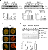
Figure 6. AKT regulates ERα protein expression.
A. ERα levels determined by western blot are reduced in C4-HI tumors treated with PD98059 or LY294002 administrated as indicated in Figure 1. However, ERα levels are more variable between tumors and not regulated by either inhibitor in C4-HD tumor samples. Top. Two representative samples of a total of six are shown in the gel. Bottom. Quantification of ERα relative to β-actin (used as a loading control) levels. n = 6 for each treatment; **:p<0.01 vs. control. B. A similar pattern of regulation of ERα protein is observed in primary cells cultured on top of Matrigel and incubated for 48 hrs with 10 µM of either of the inhibitors. Top. Two representative samples of a total of six of each group are shown in two independent gels. Bottom. Quantificaton of ERα relative to β-actin in each cell type. n = 6 for each treatment; **p<0.01 vs. control. C. Representative confocal images showing ERα determined by immunofluoresence (green). Lower levels of the protein are seen in PD98059-treated and in LY294002-treated C4-HI cells growing on Matrigel, whereas similar levels of ERα are seen in C4-HD treated and un-treated cells. Nuclei were stained red with propidium iodide. Scale bar: 30 µm. D. Top. A representative sample for each treatment is shown in a western blot. Scp2, a mouse mammary cell line, transfected with a constitutively active form of AKT1, myristoylated AKT1-Δ4-129 (Scp2Akt), displays in the same gel AKT with a typical molecular weight of 59 kDa and the myristoylated deleted variant of AKT1 (p-myrAKT) with a molecular weight of 45 kDa. The antibody used to detect total AKT recognizes only wild type AKT. E-cadherin was used as a loading control for Scp2 cells. Scp2Akt cells exhibit higher levels of ERα than Scp2 cells transfected with the vector control (vc) or Scp2 control cells. In Scp2 and Scp2vc cells, but not in Scp2Akt cells, 2 and 5 µM LY294002 downregulate p-AKT and ERα levels, whereas total AKT levels remains invariable. Bottom. Quantification of ERα relative to E-cadherin levels. n = 4 for each treatment; a,b p<0.05 Scp2Akt vs. Scp2 and Scp2vc; *p<0.05 LY294002 vs. control.
Figure 7
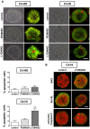
Figure 7. C4-HD tumor cells are more sensitive to steroid receptor inhibitors.
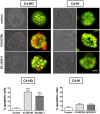
Apoptosis induced by inhibitors of ER (ICI182780) and PR (ZK230211) is higher in C4-HD than C4-HI cells. Top: Phase contrast microscopy showing representative C4-HD (left) and C4-HI (right) cell clusters in the presence of 1 µM ICI182780, 0.01 µM ZK230211 or vehicle as control. Confocal images from a fluorescence microscope of AO/EB staining show a higher number of apoptotic cells (red cells) in C4-HD cultures treated with ICI182780 or ZK230211 than in C4-HD untreated cultures. C4-HI cultures exhibit no change in cell death after endocrine treatments. Bottom. Quantification of the percentage of apoptotic C4-HD (left) and C4-HI (right) cells per cluster, of four independent experiments with ten clusters in each. Data corresponds to the mean +/− SEM. ***:p<0.001 vs. control. Scale bar: 30 µm.
Figure 8
Figure 8. Loss of endocrine resistance in isolated C4-HIR tumor cells is not prevented by 3D Matrigel.
Isolated C4-HI and C4-HIR tumor cells are sensitive to cell death in response to RU486 when cultured on plastic, and the 3D Matrigel culture system is not sufficient to revert this phenotype. A. C4-HI and C4-HIR tumors were transplanted in syngeneic mice and measured every 2 days (length and width). RU486 was inoculated s.c. at a dose of 12 mg/kg/day once the tumors were 60 mm2. C4-HI tumors (left) regress following treatment with antiprogestin, while C4-HIR tumors (right) are unresponsive. B. Fluorescent confocal images of AO/EB staining show similar number of apoptotic cells (red cells) after 48 hrs of treatment with 0.01 µM MPA and a higher number of apoptotic cells with 0.01 µM RU486 treatment. Right. Quantification of the percentage of apoptotic C4-HI (upper) and C4-HIR (lower) cells per cluster, of three independent experiments with ten clusters in each. Data corresponds to the mean +/− SEM; ***:p<0.001 vs. control. Scale bar: 100 µm.

Similar articles

Cited by

References

    1. Musgrove EA, Sutherland RL. Biological determinants of endocrine resistance in breast cancer. Nat Rev Cancer. 2009;9:631–643. - PubMed
    1. Robertson JF. Fulvestrant (Faslodex)—how to make a good drug better. Oncologist. 2007;12:774–784. - PubMed
    1. Macedo LF, Sabnis GJ, Goloubeva OG, Brodie A. Combination of anastrozole with fulvestrant in the intratumoral aromatase xenograft model. Cancer Res. 2008;68:3516–3522. - PubMed
    1. Klijn JG, de Jong FH, Bakker GH, Lamberts SW, Rodenburg CJ, et al. Antiprogestins, a new form of endocrine therapy for human breast cancer. Cancer Res. 1989;49:2851–2856. - PubMed
    1. Liang Y, Besch-Williford C, Brekken RA, Hyder SM. Progestin-dependent progression of human breast tumor xenografts: a novel model for evaluating antitumor therapeutics. Cancer Res. 2007;67:9929–9936. - PubMed

Publication types

MeSH terms