. 2010 May 26;5(5):e10830.
doi: 10.1371/journal.pone.0010830.

T cells specifically targeted to amyloid plaques enhance plaque clearance in a mouse model of Alzheimer's disease

Affiliations

T cells specifically targeted to amyloid plaques enhance plaque clearance in a mouse model of Alzheimer's disease

Yair Fisher et al. PLoS One. .

Abstract

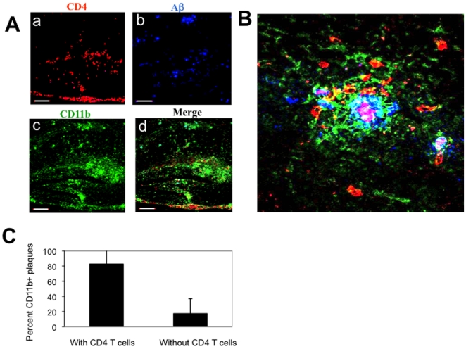
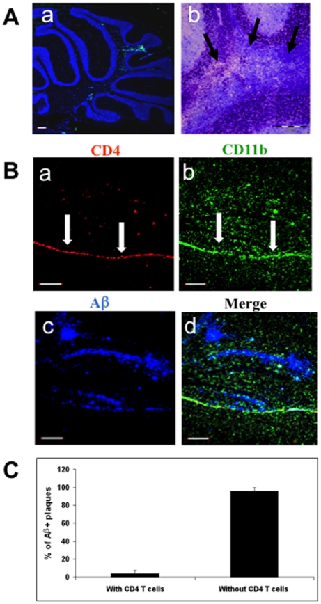
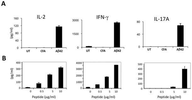
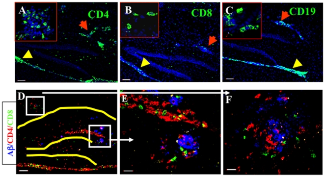
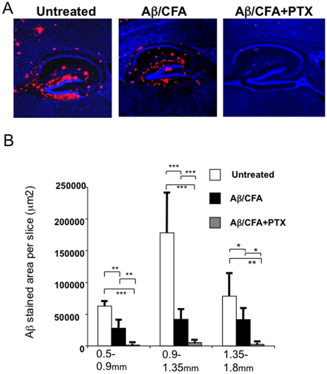
Patients with Alzheimer's disease (AD) exhibit substantial accumulation of amyloid-beta (Abeta) plaques in the brain. Here, we examine whether Abeta vaccination can facilitate the migration of T lymphocytes to specifically target Abeta plaques and consequently enhance their removal. Using a new mouse model of AD, we show that immunization with Abeta, but not with the encephalitogenic proteolipid protein (PLP), results in the accumulation of T cells at Abeta plaques in the brain. Although both Abeta-reactive and PLP-reactive T cells have a similar phenotype of Th1 cells secreting primarily IFN-gamma, the encephalitogenic T cells penetrated the spinal cord and caused experimental autoimmune encephalomyelitis (EAE), whereas Abeta T cells accumulated primarily at Abeta plaques in the brain but not the spinal cord and induced almost complete clearance of Abeta. Furthermore, while a single vaccination with Abeta resulted in upregulation of the phagocytic markers triggering receptors expressed on myeloid cells-2 (TREM2) and signal regulatory protein-beta1 (SIRPbeta1) in the brain, it caused downregulation of the proinflammatory cytokines TNF-alpha and IL-6. We thus suggest that Abeta deposits in the hippocampus area prioritize the targeting of Abeta-reactive but not PLP-reactive T cells upon vaccination. The stimulation of Abeta-reactive T cells at sites of Abeta plaques resulted in IFN-gamma-induced chemotaxis of leukocytes and therapeutic clearance of Abeta.


Conflict of interest statement

Competing Interests: The authors have declared that no competing interests exist.

Figures

Figure 1
Figure 1. Aβ immunization results in trafficking of T cells to sites of Aβ plaques in the brain parenchyma.
APP/IFN-γ Tg mice aged 9 months were immunized with Aβ and killed 19 days later. Brain sections were immunolabeled for Aβ plaques co-localized with lymphocyte subpopulations and activated microglia. (A) Brain sections derived from a representative Aβ-immunized APP/IFN-γ Tg mouse (n = 11) were immunolabeled with anti-CD4 (a, red), anti-Aβ (b, blue), and anti-CD11b (c, green) antibodies. The merged panel is shown in d. Bar represents 200 µm. (B) Three-dimensional representation of Z-stalk images taken from a representative compact Aβ plaque co-localized with CD11b+ microglia (green) and CD4 T cells (red) in the hippocampus. Bar represents 20 µm. (C) Co-localization analysis of CD4 T cells and CD11b-labeled plaques. Using Volocity 3D image analysis software (Improvision, Waltham, MA, USA), we set an intensity threshold to mark only those areas with CD11bhigh microglia, representing Aβ plaques. Columns represent percent CD11b-labeled plaques with and without co-localized CD4 T cells in each of the analyzed sections [n = 3 (3 brain sections per mouse); means ± SD; P<0.001, Student's t test].
Figure 2
Figure 2. PLP immunization results in limited T-cell occurrence at the hippocampus of APP Tg mice.
APP/IFN-γ Tg mice aged 9 months (n = 5) were immunized with PLP and killed 19 days later. (A) Brain sections were analyzed for leukocyte infiltrates as described in Materials and Methods. Sections were counterstained with TO-PRO 3 (blue). (a) Brain section showing infiltrating CD4 T cells (green) in the cerebellum. (b) Magnified area of the cerebellum stained with Luxol Fast Blue, demonstrating areas with infiltrated cells and loss of myelin (black arrows). Bar represents 200 µm. (B) Brain sections derived from a representative PLP-immunized APP/IFN-γ Tg mouse were immunolabeled with anti-CD4 (a, red), anti-CD11b (b, green), and anti-Aβ (c, blue) antibodies. The merged panel is shown in d. Bar represents 200 µm. (C) Co-localization analysis of CD4 T cells and Aβ-labeled plaques. Using Volocity 3D image analysis software (Improvision, Waltham, MA, USA), we set an intensity threshold to mark only those areas with Aβ plaques. Columns represent percent Aβplaques with and without co-localized CD4 T cells in each of the analyzed sections [n = 3 (3 brain sections per mouse); means ± SD].
Figure 3
Figure 3. Aβ or PLP vaccination of APP-Tg mice induces primarily a Th1 type of immune response.
APP/IFN-γ Tg mice aged 9–10 months were injected subcutaneously with Aβ (A) or PLP (B) emulsified in CFA in conjunction with PTX injections. Control mice were injected with CFA or left untreated (A). Mice were killed 10 days later, and popliteal lymph nodes were harvested and analyzed for cytokine secretion by ELISA as described in Materials and Methods. Lymph node derived cells were cultured without antigen or stimulated with increasing concentrations of Aβ and PLP (0.1, 1 and 10 µg/ml). Secretion of IL-2, IFN-γ, and IL-17A was measured after 24, 48, and 72 h in culture, respectively. Data obtained at 10 µg/ml Aβ are shown in A. The results shown in each case are the values obtained for three mice (means ± SD) in one representative experiment of at least three performed.
Figure 4
Figure 4. Aβ immunization results in immune-cell trafficking to the brain.
APP/IFN-γ. Tg mice aged 9 months were immunized with Aβ and killed 19 days later, and brain sections were immunolabeled as described in Materials and Methods. Lymphocyte subpopulations immunostained for CD4 (A, green), CD8 (B, green), and CD19 (C, green, with red arrows indicating infiltrates in parenchymal vessels). Yellow arrowheads indicate meningeal infiltrates. Sections were counterstained with TO-PRO 3 (blue). Bars represent 100 µm; inserts were taken with a 60× objective lens. (D) Overview of the hippocampal area (hippocampus and dentate gyrus indicated in yellow) showing CD4 (red) and CD8 (green) T cells co-localized with Aβ plaques (blue). Bar represents 100 µm. Higher magnification of Aβ plaques (blue) co-localized with CD4 (red) and CD8 (green) T cells adjacent to a parenchymal blood vessel in the dentate gyrus (E) and the CA1 (F) of the hippocampal formation. Bars represent 20 µm. Representative images from eight mice are shown.
Figure 5
Figure 5. T-cell infiltrates in the parenchyma promote efficient clearance of existing Aβ plaques.
APP/IFN-γ Tg mice aged 9 months were immunized with Aβ emulsified in CFA, Aβ emulsified in CFA in conjunction with injection of PTX, or left untreated and examined 5 weeks later for Aβ in the brain. (A) Brain sections were immunolabeled with anti-Aβ antibody (red) and counterstained with TO-PRO 3 (blue). A representative section from each group is shown. (B) The amount of Aβ in each group was quantified stereologically on 40-mm-thick sections using the Volocity 3S Image Analysis software, as described in Materials and Methods and in Supplemental data, Fig. S4. One-tailed Student's t-test revealed significant differences between the three groups of mice (n = 4 mice/group; *P<0.05, **P<0.01, ***P<0.001).

Similar articles

Cited by

References

    1. Selkoe DJ. Presenilin, Notch, and the genesis and treatment of Alzheimer's disease. Proc Natl Acad Sci U S A. 2001;98:11039–11041. - PMC - PubMed
    1. Rosenmann H, Grigoriadis N, Eldar-Levy H, Avital A, Rozenstein L, et al. A novel transgenic mouse expressing double mutant tau driven by its natural promoter exhibits tauopathy characteristics. Exp Neurol. 2008;212:71–84. - PubMed
    1. Shankar GM, Li S, Mehta TH, Garcia-Munoz A, Shepardson NE, et al. Amyloid-beta protein dimers isolated directly from Alzheimer's brains impair synaptic plasticity and memory. Nat Med 2008 - PMC - PubMed
    1. Akiyama H, Barger S, Barnum S, Bradt B, Bauer J, et al. Inflammation and Alzheimer's disease. Neurobiol Aging. 2000;21:383–421. - PMC - PubMed
    1. de Rosbo NK, Ben-Nun A. T-cell responses to myelin antigens in multiple sclerosis; relevance of the predominant autoimmune reactivity to myelin oligodendrocyte glycoprotein. J Autoimmun. 1998;11:287–299. - PubMed

Publication types

MeSH terms