. 2010 May 24;5(5):e10802.
doi: 10.1371/journal.pone.0010802.

Trafficking through COPII stabilises cell polarity and drives secretion during Drosophila epidermal differentiation

Affiliations

Trafficking through COPII stabilises cell polarity and drives secretion during Drosophila epidermal differentiation

Michaela Norum et al. PLoS One. .

Abstract

Background: The differentiation of an extracellular matrix (ECM) at the apical side of epithelial cells implies massive polarised secretion and membrane trafficking. An epithelial cell is hence engaged in coordinating secretion and cell polarity for a correct and efficient ECM formation.

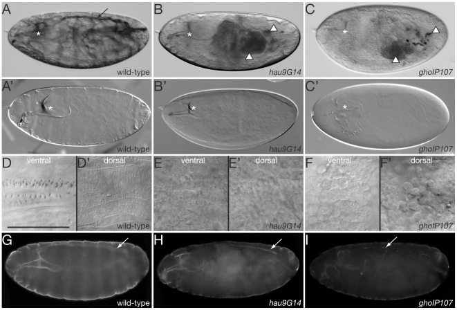
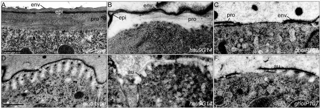
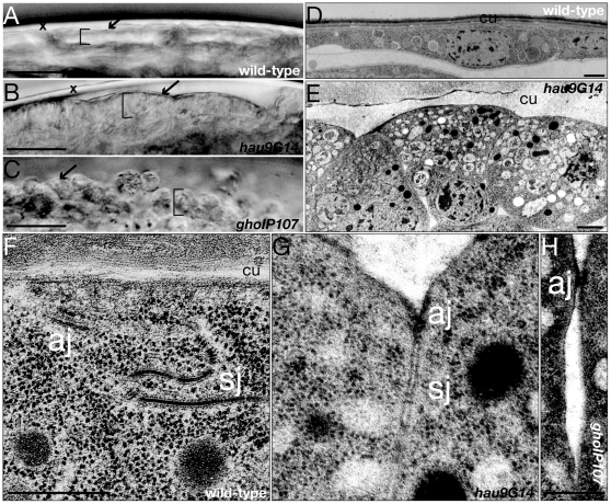
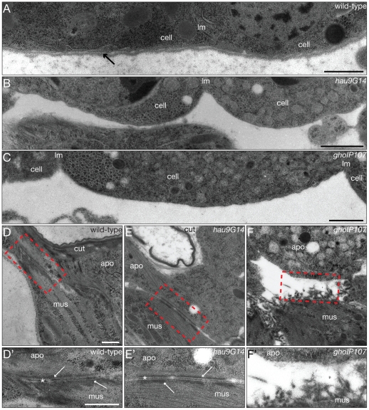
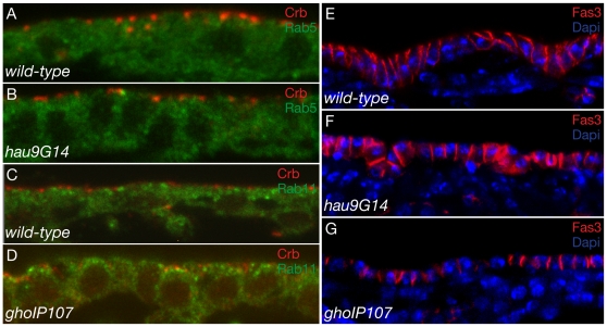
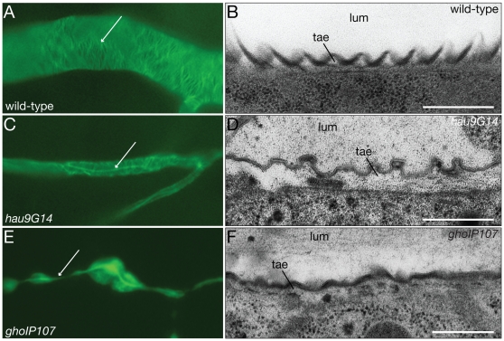
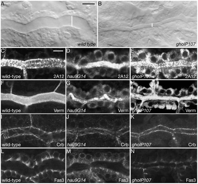
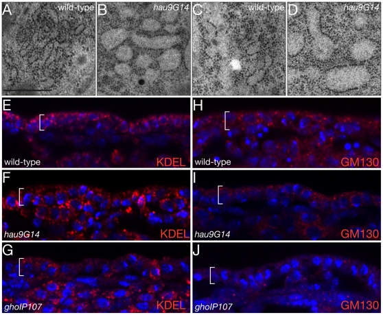
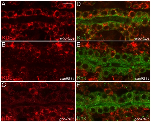
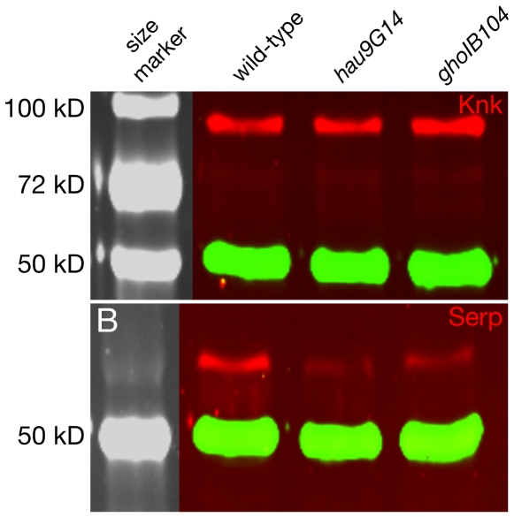
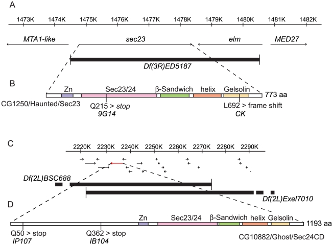
Principal findings: We are studying the molecular mechanisms that Drosophila tracheal and epidermal cells deploy to form their specific apical ECM during differentiation. In this work we demonstrate that the two genetically identified factors haunted and ghost are essential for polarity maintenance, membrane topology as well as for secretion of the tracheal luminal matrix and the cuticle. We show that they code for the Drosophila COPII vesicle-coating components Sec23 and Sec24, respectively, that organise vesicle transport from the ER to the Golgi apparatus.

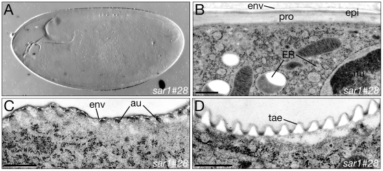
Conclusion: Taken together, epithelial differentiation during Drosophila embryogenesis is a concerted action of ECM formation, plasma membrane remodelling and maintenance of cell polarity that all three rely mainly, if not absolutely, on the canonical secretory pathway from the ER over the Golgi apparatus to the plasma membrane. Our results indicate that COPII vesicles constitute a central hub for these processes.


Conflict of interest statement

Competing Interests: The authors have declared that no competing interests exist.

Figures

Figure 1
Figure 1. Larvae carrying mutations in hau and gho have a thin and pale cuticle.
Wild-type larvae ready to hatch have a body cuticle that shrouds the inner organs (A). The head skeleton (*) and the air-filled dorsal trunk (arrow) are nevertheless discernable. By Nomarski microscopy, light refraction reveals the body cuticle of wild-type larvae within the egg case fixed in Hoyer's medium (A’). Due to a thin cuticle, the inner organs, such as the Malpighian tubules and the digestive system (arrowheads) of larvae mutant for hau and gho are well visible (B and C). Probably due to the failure to air-fill, their dorsal trunk is not identifiable at this magnification (compare to Figure 7). As seen in Hoyer's fixed hau and gho larvae, the mutant cuticle only weakly refracts light in Nomarski optics (B’ and C’). The head skeleton of these larvae seems to have a correct morphology but is less tanned. The ventral side of the wild-type larval body is decorated by belts of denticles, while more filigree hairs cover the dorsal side (D and D’). The ventral and dorsal sides of hau and gho mutant larvae, by contrast, are naked (E–F’). The surface of both mutant larvae is, however, not smooth but wrinkly, and in gho mutant larvae, epidermal cells at both sides appear to round up and leave the epithelium (see Figure 3). The epidermis, the dorsal trunk (arrow) and the head skeleton of wild-type stage 17 embryos are lined by chitin as detected with the FITC-conjugated chitin-binding probe (green, G). The chitin signal in the head skeleton and the body of hau and gho mutant stage 17 embryos is weaker than in wild-type embryos (H and I). Moreover, their dorsal trunk (arrow) is narrower than the wild-type one. (A–F’) Nomarski light microscopy of wild-type, hau and gho mutant larvae within the egg case. (G–I) Fluorescence microscopy of heat fixed stage 17 embryos. Scale bar in (D) is 50µm and applies to (D–F’).
Figure 2
Figure 2. Hau and Gho are required for full cuticle differentiation.
The wild-type larval cuticle is a stratified extracellular matrix (A). Based on the molecular composition, it is subdivided into three layers. The envelope (env) is the outermost layer, separated by the bipartite epicuticle (epi) from the innermost procuticle (pro). The cuticle of hau mutant larvae is disorganised (B). The electron-dense basal sublayer of the epicuticle often contacts the envelope and the chitin matrix has lost its tight packaging. In gho mutant larvae, the cuticle is fragmented and thin (C). Between late stage 16 and mid-stage 17 the apical plasma membrane of epidermal cells forms regular corrugations called apical undulae (au), at the tip of which chitin synthesis takes place, while secretion occurs at the valley between the corrugations (D). The epidermal cells of hau and gho mutant embryos fail to form repeated corrugations (E and F). (A–F) Electronmicrographs of ultrathinsections. Scale bar in (A) is 500nm and applies to (B) and (C). Scale bar in (D) is 500nm and applies to (E) and (F).
Figure 3
Figure 3. Hau and Gho are involved in shaping the larval epidermal cell.
The cuticle (arrow) lines the apical side of the epidermis (bracket, A). The cuticle of hau and gho mutant larvae is thinner and discontinuous (arrows, B and C). Their epidermal cells (bracket) are cuboidal (B) or round and may lose contact to their neighbours (C, see also Figure 1F,F’). The wild-type larval epidermal cell is flat with large apical surface covered by the cuticle (cu, D). Epidermal cells of hau mutant larvae are cuboidal and their lateral membranes are straight (E). Epidermal cells of gho mutant larvae display the same phenotype (not shown). The wild-type epidermal cell contacts its neighbours with its meandering lateral membrane (F). Histologically, the two obvious contact features are the subapical adherens junctions (aj) and the lateral septate junctions (sj). The lateral membranes of epidermal cells of hau mutant larvae are straight and the junctions appear less prominent (G). Epidermal cells of gho mutant larvae show a similar phenotype (not shown). Occasionally and especially in the gho mutant larval epidermis, the cell-cell contacts are lost (H). (A–C) Light-microscopy of living wild-type, hau and gho mutant larvae within the egg case (x). (D–H) Electronmicrographs of ultrathinsections. Scale bar in (A–C) is 25µm. Scale bars in (D and E) are 1 µm. Scale bar in (F) is 500 nm and applies also to (G). Scale bar in (H) is 500 nm.
Figure 4
Figure 4. Hau and Gho conduct basement membrane production.
The basal side of the wild-type larval epidermal cells (cell) separated by the lateral plasma membrane (lm) is underlain by the extracellular basement membrane (arrow, A). The basement membrane is missing in hau and gho mutant larval epidermal cells (B and C). The basal ECM (*) of wild-type larval apodemal cells (apo) is mediating the contact to muscles (mus) attached to the epidermis (D,D’). At the inner side of the apodemal cell and muscles electron-dense junctional material accumulates (arrows). The hau mutant larval apodemes have a normal-looking basal ECM, the muscular intracellular junctional material, however, is less abundant (E,E’). The gho mutant larval apodemal basal ECM is disrupted, and muscles detach from the epidermis (F,F’). No junctional material is detected at the inner side of the apodemal cell of these larvae. The red dashed rectangle in (D–F) is enlarged in (D’–F’). (A–F’) Electron micrographs of ultrathinsections. Scale bars in (A–C) are 1µm. Scale bar in (D) is 500nm and applies also to (E) and (F). Scale bar in (D’) is (500nm and applies also to (E’) and (F’).
Figure 5
Figure 5. Hau and Gho stabilise epidermal cell polarity.
The transmembrane protein Crb (red) localises to the apico-lateral region of wild-type stage 15 embryonic epidermal lateral plasma membrane (A). The small GTPase Rab5 (green) is distributed in the cytoplasm. Localisation of Crb and Rab5 is normal in hau mutant embryonic epidermal cells (B). At stage 16, Crb continues to localise to the apico-lateral region of the lateral plasma membrane (C). The small GTPase Rab11 (green) is distributed in the cytoplasm with a considerable accumulation at the apical portion of the cell. The Crb signal is detected also in the cytoplasm of gho mutant embryonic cells, while Rab11 distribution is normal (D). The lateral plasma membrane of wild-type embryos – here stage 16 - is marked by proteins like Fas3 (red) constituting the septate junctions (E). The Fas3 signal gradually decreases from the apical to the basal end of the lateral plasma membrane. The nuclei (blue) of these cells locate to the basal side of the columnar cell. In hau and gho embryonic mutant epidermal cells the Fas3 signal is homogeneously distributed along the lateral plasma membrane (F and G). The epidermal cells are cuboidal and the nuclei are lie in the middle of the cell. Images from confocal microscopy.
Figure 6
Figure 6. Hau and Gho are needed for the formation of the tracheal cuticle.
In the wild-type tracheal cuticle of the dorsal trunk and the primary branches of late stage 17 embryos, chitin is organised in a spiral running perpendicular to the length of the tube (A). Remnants of the luminal chitin are visible (arrow). These chitin cables constitute the taenidial folds (tae), which are bulges of the larval cuticle (B). At the larval stage, the lumen (lum) of the tracheal tubes does not contain any solid material. In hau late stage 17 mutant embryos, the chitin cables of the dorsal trunk and the primary branches are properly formed (C). The tracheal lumen, however, is much narrower compared to the wild-type lumen. The hau larval tracheal cuticle dilates and the taenidial folds are sloppy (D). The lumen of the hau mutant larval tracheae is not completely cleared. In gho stage 17 mutant embryos, chitin cables are largely disorganised and often absent (E). The tracheal tubes have an irregular diameter. The gho mutant larval tracheae have shallow taenidiae and their lumen fails to be cleared (F). (B,D,F) Electronmicrographs. Scale bars are 500nm. (A,C,E) Fluorescence microscopy.
Figure 7
Figure 7. Apical secretion in tracheal cells utilizes Hau and Gho function.
At early stage 17, the wild-type dorsal trunk has attained its final diameter of around 12 microns (white line, A). The early stage 17 dorsal trunks of hau and gho mutant embryos are narrower (B). During tracheal tube diameter expansion, the luminal marker 2A12 is secreted into the lumen of the tracheae (C). Most of the 2A12 signal remains within the tracheal cells of hau and gho mutant embryos during tube diameter expansion (D,E). The luminal chitin deacteylase Verm is involved in modifying the tracheal luminal chitin cable that participates in lumen diameter regulation (F). In hau and gho mutant tracheae large amounts of Verm fail to be secreted (G,H). Crb marks the apical plasma membrane in wild-type tracheal cells (I). In the tracheal cells of hau and gho mutant stage 16 embryos the localisation of Crb is unchanged (J,K). The membrane protein Fas3 lines the lateral membrane of wild-type stage 16 tracheal cells (L). In hau mutant embryos, some Fas3 signal is cytoplasmic (M). In gho mutant embryos, Fas3 localisation is as in the wild-type tracheal cells, the signal levels, however, seem to be reduced (N). Images from Confocal microscopy.
Figure 8
Figure 8. ER morphology and Golgi identity require Hau and Gho function.
The wild-type embryonic stage 17 ER in the epidermal cells is tubular (A). The ER of epidermal cells in hau mutant stage 17 embryos has, by contrast, a bloated appearance (B). Compared to the wild-type tubular ER at stage 15 (C), the dilated ER phenotype is apparent already before massive cuticle formation (D). ER residual proteins are detected by the antibody directed against the KDEL sequence. In the wild-type stage 16 embryo the KDEL antibody recognises dots in the cytoplasm (E). In the epidermis of hau and gho mutant embryos, the KDEL signal appears to be normal (F,G). The Golgi apparatus in the wild-type stage 16 epidermis is recognised by the antibody against the Golgi-specific protein GM130 and appears as dots of different sizes (H). In the hau mutant stage 16 epidermis the GM130 signal is weaker (I). The GM130 is barely detected in gho mutant stage 16 epidermal cells (J). (A–D) Electronmicroghraphs. Scale bar in (A) is 500nm and applies also to (B–D). (E–J) Images from Confocal microscopy.
Figure 9
Figure 9. Tracheal ER identity and secretion depend on Hau and Gho function.
In wild-type tracheal cells at late stage 16, the KDEL signal (red) is distributed within the cytoplasm (A,B). The membrane-associated protein Knk (green) localises to the apical plasma membrane in these cells (B). The green signal in the tracheal lumen is unspecific background. The KDEL signal in hau and gho mutant tracheal cells is strongly reduced (C–F). In both mutant tracheal cells, Knk fails to localise to the apical plasma membrane and accumulated within the cytoplasm (D,F). (A–F) Images from Confocal microscopy.
Figure 10
Figure 10. ER function is not severely affected in hau and gho mutant embryos.
The migration behaviour of the membrane-associated cuticle factor Knk (red, A) and the extracellular Serp (red, B) is normal in hau and gho mutant larvae in western blot experiments. The amount of Serp protein is reduced in the mutant protein extracts. By contrast, the amounts of Knk are comparable in mutant and wild-type protein extracts. Tubulin (green) was detected to control the amount of protein blotted. The tubulin signal allows comparing the signal intensities in wild-type and mutant protein extracts.
Figure 11
Figure 11. Molecular identification of hau.
The hau mutations were mapped to the deficiency Df(3R)ED5187 that uncovers two genes: sec23 and elm (A). Sec23 is a component of the COPII complex. The elm gene plays a role in memory formation and ethanol sensitivity, and mutations in this gene are not lethal . A nonsense mutation was identified in the sec23-coding region of the 9G14 allele, and a frame shift mutation was identified in the same coding region of the CK allele (B). Sec23 is characterised by five motifs. From the N-terminus to the C-terminus, these are the Zn binding domain, the Sec23/24 domain, which belongs to the von Willebrand factor type A (vWFA) domain family, a β-sandwich domain, a helical domain and finally a Gelsolin domain. Molecular identification of gho. The gho mutations were mapped to the interval framed by the break points of the deficiencies Df(2L)BSC688 and Df(2L)Excel7010 (C). Among the 16 genes in this region, one codes for a Sec24-like protein, CG10882 (D). In the coding region of this gene, we identified one early nonsense mutation in each allele, IB104 and IP107.
Figure 12
Figure 12. Deletion of sar1 causes a weak secretion phenotype.
Larvae having a deletion of the sar1 locus have a thin and pale cuticle (A). The three histologically distinct layers, envelope (env), epicuticle (epi) and procuticle (pro) are established (B). The ER in the epidermal cells of sar1 mutant larvae is bloated. The apical undulae (au, dotted line) of stage 16 sar1 mutant embryos are formed (C). Likewise, the taenidial folds (tae) are correctly established in sar1 deficient larvae (D). (A–D) Electron micrographs. Scale bars in (B,C,D) are 500nm.

Similar articles

Cited by

References

    1. Wickner W, Schekman R. Membrane fusion. Nat Struct Mol Biol. 2008;15:658–664. - PMC - PubMed
    1. Musch A. Microtubule organization and function in epithelial cells. Traffic. 2004;5:1–9. - PubMed
    1. Mellman I, Nelson WJ. Coordinated protein sorting, targeting and distribution in polarized cells. Nat Rev Mol Cell Biol. 2008;9:833–845. - PMC - PubMed
    1. Wodarz A, Hinz U, Engelbert M, Knust E. Expression of crumbs confers apical character on plasma membrane domains of ectodermal epithelia of Drosophila. Cell. 1995;82:67–76. - PubMed
    1. Medina E, Williams J, Klipfell E, Zarnescu D, Thomas G, et al. Crumbs interacts with moesin and beta(Heavy)-spectrin in the apical membrane skeleton of Drosophila. J Cell Biol. 2002;158:941–951. - PMC - PubMed

Publication types

MeSH terms

Substances

LinkOut - more resources