

Review
doi: 10.1007/s12350-010-9255-x.
Anatomy and physiology of coronary blood flow
Affiliations
- PMID: 20521136
- PMCID: PMC2906723
- DOI: 10.1007/s12350-010-9255-x
Item in Clipboard
Review
Anatomy and physiology of coronary blood flow
J Nucl Cardiol.
2010 Aug.
No abstract available
Figures
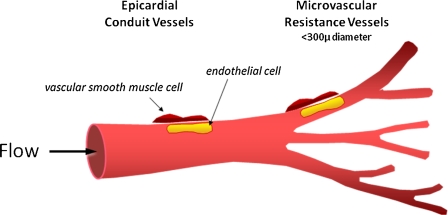
Highly schematic depiction of the main anatomic features of the coronary circulation (see text)
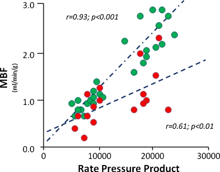
Relationship of myocardial blood flow to cardiac work as reflected by the rate pressure product. Myocardial blood flow was measured with N-13 ammonia at baseline and again during intravenous dobutamine infusion. Regional myocardial blood flows in myocardial territories supplied by nondiseased vessels are shown in green. In contrast, regional myocardial blood flows in territories subtended by diseased coronary vessels are shown in red. Adopted from Krivokapich et al
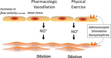
This highly simplified cartoon depicts the interaction between endothelial and vascular smooth muscle cells during pharmacologic vasodilation during physical stress
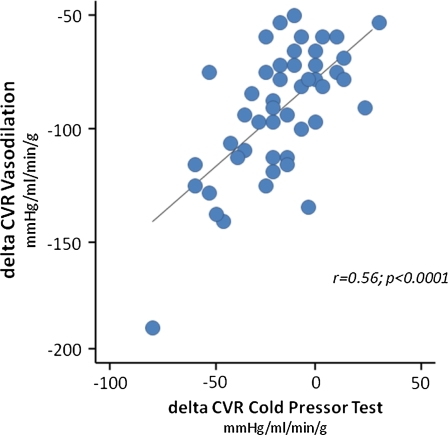
Changes in the CVR in response to cold pressor testing are compared to changes in the CVR during adenosine or dipyridamole stimulated hyperemia. Adopted from Prior et al
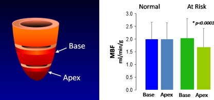
Longitudinal myocardial perfusion gradient during pharmacologically-stimulated vasodilation in normal volunteers without risk factors for coronary artery disease (blue) and in normal volunteers but with conventional risk factors. The panel on the left depicts a cartoon of the left ventricular myocardium and indicates the sites of blood flow measurements, with “base” for measurements in the left mid-ventricle and “apex” for measurements in the more apical portion of the let ventricular myocardium. Hyperemic flows recorded in these areas are depicted on the right; they are nearly identical in the two regions in the normal volunteers without risk factors but differ in individuals with risk factors with significantly lower flows in the apical than in the mid-ventricular region. Courtesy of Schindler et al from UCLA

Inverse correlation between regional myocardial blood flow during dobutamine stress and the percent coronary diameter stenosis as determined by quantitative coronary angiography. Adopted from Krivokapich et al
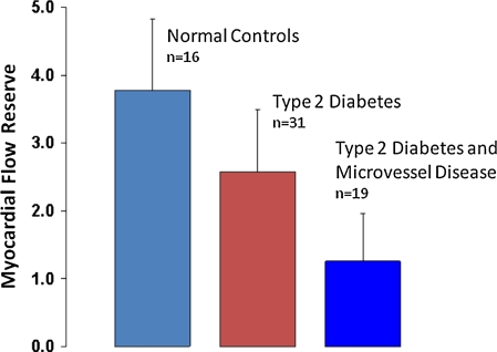
Myocardial flow reserve in patients with type 2 diabetes or in patients with type 2 diabetes and microvascular disease as compared to normal control individuals. Data taken from Yokoyama et al,,
References
-
- Bergmann SR, Fox KA, Rand AL, McElvany KD, Welch MJ, Markham J, Sobel BE. Quantification of regional myocardial blood flow in vivo with h215o. Circulation. 1984;70:724–733. - PubMed
-
- Kuhle WG, Porenta G, Huang SC, Buxton D, Gambhir SS, Hansen H, Phelps ME, Schelbert HR. Quantification of regional myocardial blood flow using 13n-ammonia and reoriented dynamic positron emission tomographic imaging. Circulation. 1992;86:1004–1017. - PubMed
-
- Lautamaki R, George RT, Kitagawa K, Higuchi T, Merrill J, Voicu C, DiPaula A, Nekolla SG, Lima JA, Lardo AC, Bengel FM. Rubidium-82 pet-ct for quantitative assessment of myocardial blood flow: Validation in a canine model of coronary artery stenosis. Eur J Nucl Med Mol Imaging. 2009;36:576–586. doi: 10.1007/s00259-008-0972-1. - DOI - PubMed
-
- Muzik O, Beanlands RS, Hutchins GD, Mangner TJ, Nguyen N, Schwaiger M. Validation of nitrogen-13-ammonia tracer kinetic model for quantification of myocardial blood flow using pet. J Nucl Med. 1993;34:83–91. - PubMed
