Multi-walled carbon nanotubes induce cytotoxicity, genotoxicity and apoptosis in normal human dermal fibroblast cells
- PMID: 20521388
- PMCID: PMC2902968
Multi-walled carbon nanotubes induce cytotoxicity, genotoxicity and apoptosis in normal human dermal fibroblast cells
Abstract
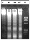
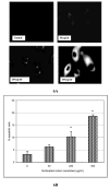
Multi-walled carbon nanotubes (MWCNT) have won enormous popularity in nanotechnology. Due to their unusual, one dimensional, hollow nanostructure and unique physicochemical properties they are highly desirable for use within the commercial, environmental and medical sectors. Despite their wide application, little information is known concerning their impact on human health and the environment. While nanotechnology looms large with commercial promise and potential benefit, an equally large issue is the evaluation of potential effects on humans and other biological systems. Our research is focused on cellular response to purified MWCNT in normal human dermal fibroblast cells (NHDF). Three doses (40, 200, 400 microg/mL) of MWCNT and control (tween-80+0.9% saline) were used in this study. Following exposure to MWCNT, cytotoxicity, genotoxicity and apoptosis assays were performed using standard protocols. Our results demonstrated a dose-dependent toxicity with MWCNT. It was found to be toxic and induced massive loss of cell viability through DNA damage and programmed cell-death of all doses compared to control. Our results demonstrate that carbon nanotubes indeed can be very toxic at sufficiently high concentrations and that careful monitoring of toxicity studies is essential for risk assessment.
Figures


Similar articles
-
Evaluation of cell viability, DNA damage, and cell death in normal human dermal fibroblast cells induced by functionalized multiwalled carbon nanotube.Mol Cell Biochem. 2010 May;338(1-2):225-32. doi: 10.1007/s11010-009-0356-2. Epub 2009 Dec 17. Mol Cell Biochem. 2010. PMID: 20016928
-
Effects of nitrogen-doped multi-walled carbon nanotubes compared to pristine multi-walled carbon nanotubes on human small airway epithelial cells.Toxicology. 2015 Jul 3;333:25-36. doi: 10.1016/j.tox.2015.03.008. Epub 2015 Mar 20. Toxicology. 2015. PMID: 25797581 Free PMC article.
-
Characterization of genotoxic response to 15 multiwalled carbon nanotubes with variable physicochemical properties including surface functionalizations in the FE1-Muta(TM) mouse lung epithelial cell line.Environ Mol Mutagen. 2015 Mar;56(2):183-203. doi: 10.1002/em.21922. Epub 2014 Nov 12. Environ Mol Mutagen. 2015. PMID: 25393212
-
Weight of evidence analysis for assessing the genotoxic potential of carbon nanotubes.Crit Rev Toxicol. 2017 Nov;47(10):867-884. doi: 10.1080/10408444.2017.1367755. Epub 2017 Sep 22. Crit Rev Toxicol. 2017. PMID: 28937307 Review.
-
Genotoxicity of multi-walled carbon nanotube reference materials in mammalian cells and animals.Mutat Res Rev Mutat Res. 2021 Jul-Dec;788:108393. doi: 10.1016/j.mrrev.2021.108393. Epub 2021 Aug 21. Mutat Res Rev Mutat Res. 2021. PMID: 34893158 Review.
Cited by
-
In vitro drug release characteristic and cytotoxic activity of silibinin-loaded single walled carbon nanotubes functionalized with biocompatible polymers.Chem Cent J. 2016 Dec 12;10:81. doi: 10.1186/s13065-016-0228-2. eCollection 2016. Chem Cent J. 2016. PMID: 28028386 Free PMC article.
-
Mechanisms of nanoparticle-induced oxidative stress and toxicity.Biomed Res Int. 2013;2013:942916. doi: 10.1155/2013/942916. Epub 2013 Aug 20. Biomed Res Int. 2013. PMID: 24027766 Free PMC article.
-
Proteomic analysis of cellular response induced by multi-walled carbon nanotubes exposure in A549 cells.PLoS One. 2014 Jan 14;9(1):e84974. doi: 10.1371/journal.pone.0084974. eCollection 2014. PLoS One. 2014. PMID: 24454774 Free PMC article.
-
In vitro toxicity of carbon nanotubes: a systematic review.RSC Adv. 2022 May 31;12(25):16235-16256. doi: 10.1039/d2ra02519a. eCollection 2022 May 23. RSC Adv. 2022. PMID: 35733671 Free PMC article. Review.
-
Multi-walled carbon nanotube directed gene and protein expression in cultured human aortic endothelial cells is influenced by suspension medium.Toxicology. 2012 Dec 16;302(2-3):114-22. doi: 10.1016/j.tox.2012.09.008. Epub 2012 Sep 28. Toxicology. 2012. PMID: 23026733 Free PMC article.
References
-
- Iijima S. Helical microtubules of graphitic carbon. Nature. 1991;354:56–59.
-
- Endo M, Strano MS, Ajayan PM. Potential applications of carbon nanotubes. Carbon Nanotubes. 2008;111:13–61.
-
- Maynard AD, Baron PA, Foley M, Shvedova AA, Kisin ER, Castranova V. Exposure to carbon nanotube material: aerosol release during the handling of unrefined single-walled carbon nanotube material. J Toxicol & Environ Health. 2004;A67(1):87–107. - PubMed
-
- Ball P. Roll up for the revolution. Nature. 2001;414:142–144. - PubMed
-
- Colvin VL. The potential environmental impact of engineered nanomaterials. Nat Biotechnol. 2003;21(10):1166–70. - PubMed
Publication types
MeSH terms
Substances
Grants and funding
LinkOut - more resources
Full Text Sources
