. 2010 Apr;41(2):61-7.
doi: 10.1177/155005941004100204.

Changing views of basal ganglia circuits and circuit disorders

Affiliations

Changing views of basal ganglia circuits and circuit disorders

Mahlon DeLong et al. Clin EEG Neurosci. 2010 Apr.

Abstract

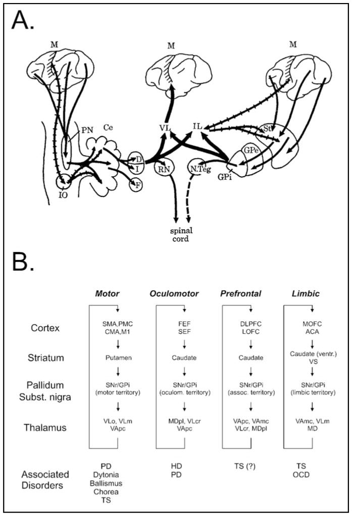
The basal ganglia (BG) have long been considered to play an important role in the control of movement and the pathophysiology of movement disorders, such as Parkinson's disease (PD). Studies over the past decades have considerably broadened this view, indicating that the BG participate in multiple, parallel, largely segregated, cortico-subcortical reentrant pathways involving motor, associative and limbic functions. Research has shown that dysfunction within individual circuits is associated not only with movement disorders, but also with neuropsychiatric disorders. Accordingly, a number of movement disorders and neuropsychiatric disorders such as obsessive compulsive disorder and Tourette's syndrome are viewed as "circuit disorders." We here discuss the changes in our current understanding of the anatomic and functional organization of BG circuits and related circuit disorders.


Conflict of interest statement

DISCLOSURE AND CONFLICT OF INTEREST

M. DeLong and T. Wichmann have no conflicts of interest in relation to this article.

Figures

Figure 1
Figure 1
Changing views of the organization of the BG and their integration in the control of motor and non-motor functions. A. View of the BG as developed by Kemp and Powell in the 1970s. Multiple motor and non-motor cortical inputs converge in the BG. BG and cerebellum interact to regulate motor cortical functions. B. Current view of the circuit anatomy of the cortex-basal ganglia-thalamocortical circuits. Motor and non-motor circuits are segregated throughout their subcortical course, involving specific territories in the BG and associated areas of thalamus and cortex. Abnormal activity in specific circuits gives rise to “circuit disorders.” Abbreviations: ACA, anterior cingulate area; CMA, cingulate motor area; DLPFC, dorsolateral prefrontal cortex; FEF, frontal eye fields; LOFC, lateral orbitofrontal cortex; M1, primary motor cortex; MD, mediodorsal nucleus of the thalamus; MDpl, mediodorsal nucleus of thalamus, pars lateralis; MOFC, medial orbitofrontal cortex; PMC, premotor cortex; SMA, supplementary motor area; SEF, supplementary eye field; VApc, ventral anterior nucleus of thalamus, pars parvocellularis; VAmc, ventral anterior nucleus of thalamus, pars magnocellularis; VLm, ventrolateral nucleus of thalamus, pars medialis; VLo, ventrolateral nucleus of thalamus, pars oralis; VLcr, ventrolateral nucleus of thalamus, pars caudalis, rostral division. See text for other abbreviations. Figure 1A reproduced, with permission, from [4]. Figure 1B reproduced, with permission, from [1].
Figure 2
Figure 2
Motor circuit of the BG. Black arrows indicate inhibitory connections; gray arrows indicate excitatory connections. The thickness of the arrows corresponds to their presumed activity. Abbreviations: CM, centromedian nucleus of thalamus; CMA, cingulate motor area; Dir., direct pathway; Hyperdir., hyperdirect pathway; D1, D2, dopamine receptor subtypes; Indir., indirect pathway; M1, primary motor cortex; PMC, premotor cortex; SMA, supplementary motor area; VA, ventral anterior nucleus of thalamus; VL, ventrolateral nucleus of thalamus. See text for other abbreviations. Reproduced, with permission, from [74].

References

    1. Wichmann T, DeLong MR. Deep brain stimulation for neurologic and neuropsychiatric disorders. Neuron. 2006;52:197–204. - PubMed
    1. Wichmann T, DeLong MR. Anatomy and physiology of the basal ganglia: relevance to Parkinson’s disease and related disorders. In: Koller W, Melamed E, editors. Handbook Clinical Neurology. Vol. 83. New York, NY: Elsevier; 2007. pp. 1–18. - PubMed
    1. DeLong M, Wichmann T. Update on models of basal ganglia function and dysfunction. Parkinsonism Relat Disord. 2009;15 (suppl 3):S237–240. - PMC - PubMed
    1. Kemp JM, Powell TPS. The connexions of the striatum and globus pallidus: synthesis and speculation. Philosophical Transactions of the Royal Society of London - Series B: Biol Sciences. 1971;262:441–457. - PubMed
    1. Alexander GE, DeLong MR, Strick PL. Parallel organization of functionally segregated circuits linking basal ganglia and cortex. Annu Rev Neurosci. 1986;9:357–381. - PubMed

MeSH terms