Review
. 2010 Sep 3;285(36):27525-31.
doi: 10.1074/jbc.R110.135970. Epub 2010 Jun 3.

Thiazolyl peptide antibiotic biosynthesis: a cascade of post-translational modifications on ribosomal nascent proteins

Affiliations
Review

Thiazolyl peptide antibiotic biosynthesis: a cascade of post-translational modifications on ribosomal nascent proteins

Christopher T Walsh et al. J Biol Chem. .

Abstract

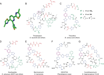
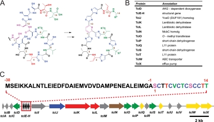
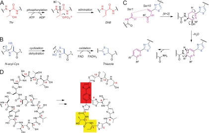
Antibiotics of the thiocillin, GE2270A, and thiostrepton class, which block steps in bacterial protein synthesis, contain a trithiazolyl (tetrahydro)pyridine core that provides the architectural constraints for high affinity binding to either the 50 S ribosomal subunit or elongation factor Tu. These mature antibiotic scaffolds arise from a cascade of post-translational modifications on 50-60-residue prepeptide precursors that trim away the N-terminal leader sequences (approximately 40 residues) while the C-terminal 14-18 residues are converted into the mature scaffold. In the producing microbes, the genes encoding the prepeptide open reading frames are flanked in biosynthetic clusters by genes encoding post-translational modification enzymes that carry out lantibiotic-type dehydrations of Ser and Thr residues to dehydroamino acid side chains, cyclodehydration and oxidation of cysteines to thiazoles, and condensation of two dehydroalanine residues en route to the (tetrahydro)pyridine core. The trithiazolyl pyridine framework thus arises from post-translational modification of the peptide backbone of three Cys and two Ser residues of the prepeptide.


Figures

FIGURE 1.
FIGURE 1.
Molecular structures of thiopeptide antibiotics. A, three-dimensional rendering of the thiocillin core depicting the propeller-like architecture resulting from macrocyclization. B, molecular structure of the eight members of the thiocillin family produced by B. cereus containing a trithiazolyl pyridine core. C, molecular structure of thiostrepton produced by Streptomyces laurentii and containing a trithiazolyl tetrahydropyridine core. Note the presence of the upstream sequence, attached to the tetrahydropyridine at the same carbon as thiazole 2, which constitutes part of the second macrocyclic loop. D, nosiheptide contains a second macrocycle consisting of an indolic acid attached between the side chains of Cys-10 and Glu-8 of the primary macrocycle. E, the macrocycle of berninamycin is extended by nine bonds between connections with the central pyridine compared with thiocillin and nosiheptide (35 bonds versus 26 bonds, respectively). F, GE2270A also has an elongated macrocycle compared with thiocillin, with 29 bonds between pyridine connections. G, the second macrocycle of cyclothiazomycin is generated by conjugation of the C-terminal segment to the side chain of a primary macrocycle amino acid.
FIGURE 2.
FIGURE 2.
Thiopeptide antibiotics bound to their targets in translation elongation. A, crystal structure of nosiheptide bound to the 50 S ribosomal subunit in a cleft formed by protein L11 (green) and the 23 S rRNA (gray). The central pyridine (pink) and thiazoles (blue) of nosiheptide are highlighted (Protein Data Bank code 2ZJP (57)). B, overlay of crystal structures of EF-Tu binding Phe-tRNA and GE2270A independently. This figure was adapted from Ref. . For clarity, only domain 2 has been aligned; domains 1 and 3 (gray), domain 2 (green), and guanosine 5-(β,γ-imido)triphosphate (GDPNP; red) correspond to EF-Tu bound to GE2270A (Protein Data Bank code 2C77 (68)). Domain 2 (blue) corresponds to EF-Tu bound to Phe-tRNA (orange) (Protein Data Bank code 1TTT (66)).
FIGURE 3.
FIGURE 3.
Gene cluster responsible for biosynthesis of the thiocillin family of thiopeptides. A, predicted deconvolution of the mature thiocillin scaffold to generate a 14-amino acid precursor that could yield thiocillin through post-translational modifications. The resulting amino acid sequence is SCTTCVCTCSCCTT. B, list of genes and their purported functions responsible for thiocillin biosynthesis. C, graphical depiction of the thiocillin biosynthetic gene cluster. Four identical copies of a gene were identified that encode a 52-amino acid peptide containing the predicted 14-amino acid precursor of thiocillin.
FIGURE 4.
FIGURE 4.
Mechanisms and essential residues involved in maturation of thiopeptide compounds. A, Dha and Dhb are formed by a two-step mechanism involving phosphorylation of the Ser or Thr side chain. Base abstraction of the β-carbon proton results in formation of the sp2 center and elimination of the phosphate. B, thiazole formation begins with attack of a Cys thiol on the carbonyl of the previous amino acid residue. Rearrangement results in dehydration producing thiazoline, which is then oxidized to thiazole in an FAD-dependent mechanism. C, formation of the central pyridine ring of thiocillin is thought to proceed by [4 + 2] cycloaddition of the Dha residues derived from Ser-1 and Ser-10. Subsequent dehydration and elimination of the leader peptide by scission of the C–N bond complete pyridine formation. D, amino acids Ser-1 and Ser-10, responsible for formation of the central pyridine of thiocillin (red), are essential for production of thiocillin. Substitutions preventing thiazole formation from Cys-11 (red) completely inhibit production of thiocillin, suggesting that Cys-11 is crucial in the maturation of the thiocillin scaffold. Although substitutions at Thr-3 and Dhb-4 (yellow) should not alter the overall architecture of the macrocycle, alanine substitutions at these positions result in loss of antibiotic activity, consistent with their purported role in ribosome recognition and binding.

References

    1. Sattely E. S., Fischbach M. A., Walsh C. T. (2008) Nat. Prod. Rep. 25, 757–793 - PubMed
    1. Walsh C. T., Fischbach M. A. (2010) J. Am. Chem. Soc. 132, 2469–2493 - PMC - PubMed
    1. Cane D. E. (2010) J. Biol. Chem. 285, 27517–27523 - PMC - PubMed
    1. Pickens L. B., Tang Y. (2010) J. Biol. Chem. 285, 27509–27515 - PMC - PubMed
    1. Fischbach M. A., Walsh C. T. (2006) Chem. Rev. 106, 3468–3496 - PubMed

Publication types

MeSH terms