

Biochemical and genetic insights into asukamycin biosynthesis
- PMID: 20522559
- PMCID: PMC2915727
- DOI: 10.1074/jbc.M110.128850
Biochemical and genetic insights into asukamycin biosynthesis
Abstract
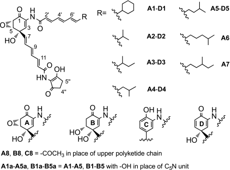
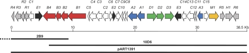
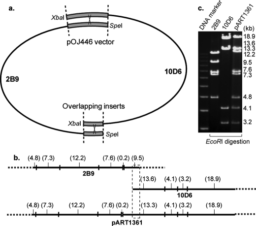
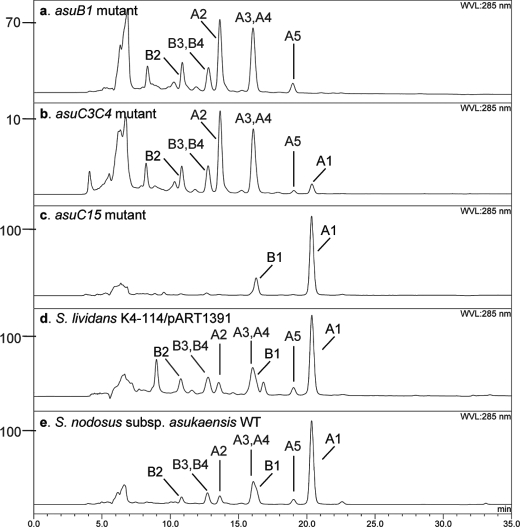
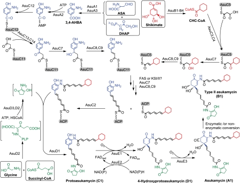
Asukamycin, a member of the manumycin family metabolites, is an antimicrobial and potential antitumor agent isolated from Streptomyces nodosus subsp. asukaensis. The entire asukamycin biosynthetic gene cluster was cloned, assembled, and expressed heterologously in Streptomyces lividans. Bioinformatic analysis and mutagenesis studies elucidated the biosynthetic pathway at the genetic and biochemical level. Four gene sets, asuA-D, govern the formation and assembly of the asukamycin building blocks: a 3-amino-4-hydroxybenzoic acid core component, a cyclohexane ring, two triene polyketide chains, and a 2-amino-3-hydroxycyclopent-2-enone moiety to form the intermediate protoasukamycin. AsuE1 and AsuE2 catalyze the conversion of protoasukamycin to 4-hydroxyprotoasukamycin, which is epoxidized at C5-C6 by AsuE3 to the final product, asukamycin. Branched acyl CoA starter units, derived from Val, Leu, and Ile, can be incorporated by the actions of the polyketide synthase III (KSIII) AsuC3/C4 as well as the cellular fatty acid synthase FabH to produce the asukamycin congeners A2-A7. In addition, the type II thioesterase AsuC15 limits the cellular level of omega-cyclohexyl fatty acids and likely maintains homeostasis of the cellular membrane.
Figures
References
-
- Sattler I., Thiericke R., Zeeck A. (1998) Nat. Prod. Rep. 15, 221–240 - PubMed
-
- Shipley P. R., Donnelly C. C., Le C. H., Bernauer A. D., Klegeris A. (2009) Int. J. Mol. Med. 24, 711–715 - PubMed
-
- Bernier M., Kwon Y. K., Pandey S. K., Zhu T. N., Zhao R. J., Maciuk A., He H. J., Decabo R., Kole S. (2006) J. Biol. Chem. 281, 2551–2561 - PubMed
-
- Zheng Z. H., Dong Y. S., Zhang H., Lu X. H., Ren X., Zhao G., He J. G., Si S. Y. (2007) J. Enzyme Inhib. Med. Chem. 22, 43–49 - PubMed
-
- Kakinuma K., Ikekawa N., Nakagawa A., Omura S. (1979) J. Am. Chem. Soc. 101, 3402–3404
Publication types
MeSH terms
Substances
Grants and funding
LinkOut - more resources
Full Text Sources
Molecular Biology Databases
Miscellaneous
