. 2010 Aug;59(8):1948-56.
doi: 10.2337/db09-1010. Epub 2010 Jun 3.

Mesenchymal bone morphogenetic protein signaling is required for normal pancreas development

Affiliations

Mesenchymal bone morphogenetic protein signaling is required for normal pancreas development

Jonas Ahnfelt-Rønne et al. Diabetes. 2010 Aug.

Abstract

Objective: Pancreas organogenesis is orchestrated by interactions between the epithelium and the mesenchyme, but these interactions are not completely understood. Here we investigated a role for bone morphogenetic protein (BMP) signaling within the pancreas mesenchyme and found it to be required for the normal development of the mesenchyme as well as for the pancreatic epithelium.

Research design and methods: We analyzed active BMP signaling by immunostaining for phospho-Smad1,5,8 and tested whether pancreas development was affected by BMP inhibition after expression of Noggin and dominant negative BMP receptors in chicken and mouse pancreas.

Results: Endogenous BMP signaling is confined to the mesenchyme in the early pancreas and inhibition of BMP signaling results in severe pancreatic hypoplasia with reduced epithelial branching. Notably, we also observed an excessive endocrine differentiation when mesenchymal BMP signaling is blocked, presumably secondary to defective mesenchyme to epithelium signaling.

Conclusions: We conclude that BMP signaling plays a previously unsuspected role in the mesenchyme, required for normal development of the mesenchyme as well as for the epithelium.


Figures

FIG. 1.
FIG. 1.
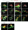
pSmad1,5,8 immunoreactivity in the embryonic chicken and mouse pancreas. pSmad1,5,8 stainings on e2, e3, and e4 (HH st.12, 18, and 21) chicken embryos (A–J), e9.5 mouse embryo (K–L), and e11.5-dissected mouse gut (M–N). A–D: e2 embryo stained in whole mount as indicated. Images are either projections of confocal z-stacks of thick vibratome sections (A–C) or an optical section (D). Boxed area in B is shown in higher magnification in C and D. A: Projected image from a z-stack showing pSmad1,5,8 reactivity in the ventral forgut endoderm (arrows) and dorsal neural tube (arrowhead). B: Projected image at the presumptive pancreas level. Note pSmad1,5,8 reactivity in the lateral plate mesoderm and the underlying endoderm that appears yellow because of coexpression with Foxa2 (arrow). C and D: pSmad1,5,8 reactivity can be observed in the endothelial cells of the dorsal aortas and in endoderm lateral to the Nkx6.1 expression domain (arrowhead). E and F: A section of an e3 pancreas stained as indicated (shown with and without Nkx6.1). Note the pSmad1,5,8 reactivity in the mesenchyme and the absence of staining in the pancreas epithelium (arrows point to the dorsal pancreas). G: Composite of two images showing the dorsal and ventral pancreas and the liver; pSmad1,5,8 can be detected in the pancreas mesenchyme and in mesenchymal and epithelial compartments of the liver. Note the sharp boundary of pSmad1,5,8 in the bile duct (arrows). H–J: Another section from the same pancreas showing absence of pSmad1,5,8 immunoreactivity in the Nkx6.1-positive pancreatic epithelium and β-cells (recognizable by their morphology, arrowhead). K and L: Optical sections from a z-stack of an e9.5 mouse stained in whole mount; pSmad1,5,8 can be detected in the dorsal aorta and intersomitic vessels and in mesenchymal cells in close contact with the ventral pancreas epithelium. M and N: Optical sections from an e11.5 mouse dissected gut. Note the mesenchymal pSmad1,5,8 signal. Dashed outline is shown without Pdx1 in the inset. *marks an unspecific luminal signal from antibody trapping. a, aorta; dp, dorsal pancreas; duo, duodenum, v, right omphalomesenteric vein; vp, ventral pancreas. (A high-quality digital representation of this figure is available in the online issue.)
FIG. 2.
FIG. 2.
Expression patterns of BMPs in the embryonic chicken pancreas—mRNA ISH, showing the expression of Bmp4 and Bmp7 at the level of the posterior stomach (in A and C, arrows point at the dorsal aorta. Arrowheads in A point to mesonephros, and arrowheads in C point to expression in the stomach mesenchyme) and the pancreas (B and D) at HH. st. 18. Note that the mesenchymal expression of Bmp4 and Bmp7 in the pancreas mesenchyme (arrows, B and D). l, liver; p, pancreas; st. stomach. (A high-quality digital representation of this figure is available in the online issue.)
FIG. 3.
FIG. 3.
Ectopic expression of Noggin in the chicken pancreas epithelium leads to loss of pSmad1,5,8 immunoreactivity in the mesenchyme. Sectioned pancreata immunolabeled as indicated, 24 h (A–D) and 48 h (E–H) after electroporation. GFP expression is showing transfected cells. Nkx6.1 is labeling the pancreas. Note the loss of pSMAD1,5,8 in the pancreas mesenchyme around the Noggin transfected area compared with controls. H: Arrowheads point to pSmad1,5,8 signals maintained at a distance from the electroporated area. dp, dorsal pancreas; vp, ventral pancreas. (A high-quality digital representation of this figure is available in the online issue.)
FIG. 4.
FIG. 4.
Ectopic Noggin expression in the pancreas results in poor epithelial branching, precocious endocrine differentiation. Electroporated pancreata immunofluorescently stained in whole mount as indicated. A–D: At 24 h after electroporation with control plasmid (A and B) and Noggin (C and D). Projected images from z-stacks show correct specification of the dorsal and ventral pancreas under both conditions. B and D are identical to A and C, except that the GFP signal was removed to show the distribution of pancreatic cell types. E and F: Forty-eight hours after electroporation with control plasmid (E and F) and Noggin (G and H). E and G: Projected images from a z-stack. F and H: Optical sections from the stack. In the control, primary branchpoint in the epithelium can be observed (F, arrowheads) and endocrine cells are restricted to the dorsal pancreas. The dorsal pancreas is completely unbranched after Noggin electroporation and extends as a tubular structure anteriorly along the stomach (arrowheads in G and H). Numerous endocrine cells can be seen in the ventral pancreas (arrow in G and H). I–L: Seventy-two hours after electroporation with control plasmid (I and J) and Noggin (K and L). I and K: Projected images from a z-stack. J and L: Optical sections from the stack. Branching is more pronounced in the controls after 72 h (J, arrowheads), and endocrine cells are still confined to the dorsal pancreas. In Noggin electroporated pancreata, almost no branching has occurred. A large cluster of pancreatic endocrine cells can be found detached from the pancreas in the stomach mesenchyme (K and L, arrowhead). In the small remaining ventral pancreas, numerous endocrine cells can be observed (K and L, arrow). M: Less frequently observed phenotype 48 h after noggin electroporation—the dorsal pancreas is almost entirely converted into a large endocrine cell mass. N: Electroporation with dnALK3 has no effect on the normal pancreas development 48 h after electroporation. See Table 1 for quantifications. dp, dorsal pancreas; vp, ventral pancreas. (A high-quality digital representation of this figure is available in the online issue.)
FIG. 5.
FIG. 5.
Pancreatic cell types can be observed in the stomach mesenchyme and exocrine differentiation is reduced 48 h after electroporation with Noggin. A and B are adjacent sections at the level of the stomach in a Noggin electroporated embryo showing Nkx6.1 mRNA expression and insulin and glucagon protein expression, respectively. Large clusters of pancreatic cells can be found in the stomach mesenchyme, and both glucagon and insulin are readily found (A and B). C and D: Sections of the pancreas of control (C) and Noggin (D) electroprated embryos imunoflurescently labeled for Amylase and showing GFP expression in electroporated cells. in D inset: The boxed area is without GFP. Note the absence of amylase-positive cells in the Noggin-electroporated pancreas. (A high-quality digital representation of this figure is available in the online issue.)
FIG. 6.
FIG. 6.
Cell autonomous BMP signaling inhibition in the mouse pancreas mesenchyme results in increased β-cell differentiation. Mouse pancreas organ cultures explanted and transduced at e12.5 (A–K) or 10.5 (L–N) with lentiviral constructs as indicated. A–C: Stereomicrographs after 3 days in culture. D–F: Stereomicrographs of three different explants of each condition after 7 days in culture. G and H: Explants transduced with a control EGFP construct, immunflurescently stained for E-cadherin (red) and showing EGFP with or without DAPI nuclear counterstain. Note how GFP expression is restricted to the mesenchyme 3 and 7 days after transduction. I–K: Explants transduced as indicated and cultured for 7 days. Immunostained for amylase (green), insulin (red), and nuclei (DAPI, blue). L–N: Explants transduced at e10.5 and cultured for 8 days, then stained for amylase and insulin with DAPI nuclear counter stain. See Table 2 for quantifications. (A high-quality digital representation of this figure is available in the online issue.)
FIG. 7.
FIG. 7.
Electroporation with Noggin results in persistence of a Bapx1-positive pancreas mesenchyme on the right side of the pancreas 48 h after electroporation. mRNA in situ hybridization on electroporated embryos 48 h after electroporation. A, C, E and B, D, F are serial sections from the same embryos, G–J are serial sections from other embryos. Nkx6.1 staining shows the pancreas epithelium (A and B). Pitx2 and Bapx1 are normally coexpressed on the left side of the pancreas in control embryos (arrows, C and E). However, in Noggin-electroporated embryos, Bapx1 expression becomes symmetrically expressed on both sides of the pancreas (arrows, F), whereas Pitx2 remains left sided (arrow, D). Most of the unbranced dorsal pancreas in Noggin electroporated embryos does not express Hes1 (G and H), but the duodenum maintains Hes1 expression. dp, dorsal pancreas; duo, duodenum; vp, ventral pancreas.
FIG. 8.
FIG. 8.
Vascular/pancreatic development in electroporated embryos 48 h after electroporation. Embryos perfused with a fluorescently-tagged Lens Culinaris Agglutinin (labeling the vascular endothelium) and subjected to whole-mount immunostaing for Nkx6.1 and GFP. A,B and E,F show projected z-stacks from lateral views at two different magnifications. C and G: Projected images from transverse thick vibratome sections from the same embryos (indicated by dashed lines in A and E). Note how the dorsal aorta is well separated from the pancreas in the control and how the dorsal pancreas curls around the omphalomesenteric vein. In Noggin-electroporated embryos, the omphalomesenteric vein is consistently found ventrally to the pancreas, and in some of the embryos, the paired dorsal aortas fail to fuse. The bisymmetrical mesenchyme is indicated by dashed lines in F. (The contrast was temporarily enhanced in the image to allow the drawing based on a background signal. The mesenchyme is not readily observable at the published contrast setting). D and H: Schematic drawing summarizing the relationship between the gut and pancreas (green) and the omphalomesenteric vein (red). D is drawn similar to a study done by Romanoff (33). a, aorta; dp, dorsal pancreas; duo, duodenum; dv, ductus venosus; li, liver; ov, omphalomesenteric vein; vp, ventral pancreas. (A high-quality digital representation of this figure is available in the online issue.)

References

    1. Jorgensen MC, Ahnfelt-Ronne J, Hald J, Madsen OD, Serup P, Hecksher-Sorensen J: An illustrated review of early pancreas development in the mouse. Endocr Rev 2007;28:685–705 - PubMed
    1. Hebrok M, Kim SK, Melton DA: Notochord repression of endodermal Sonic hedgehog permits pancreas development. Genes Dev 1998;12:1705–1713 - PMC - PubMed
    1. Kumar M, Jordan N, Melton D, Grapin-Botton A: Signals from lateral plate mesoderm instruct endoderm toward a pancreatic fate. Dev Biol 2003;259:109–122 - PubMed
    1. Lammert E, Cleaver O, Melton D: Induction of pancreatic differentiation by signals from blood vessels. Science 2001;294:564–567 - PubMed
    1. Kim SK, Hebrok M, Melton DA: Pancreas development in the chick embryo. Cold Spring Harb Symp Quant Biol 1997;62:377–383 - PubMed

Publication types

MeSH terms