. 2010 Jul;177(1):311-24.
doi: 10.2353/ajpath.2010.090915. Epub 2010 Jun 3.

Accelerated lipofuscinosis and ubiquitination in granulin knockout mice suggest a role for progranulin in successful aging

Affiliations

Accelerated lipofuscinosis and ubiquitination in granulin knockout mice suggest a role for progranulin in successful aging

Zeshan Ahmed et al. Am J Pathol. 2010 Jul.

Erratum in

  • Am J Pathol. 2010 Oct;177(4):2146. Wuertzer, Charles A [added]

Abstract

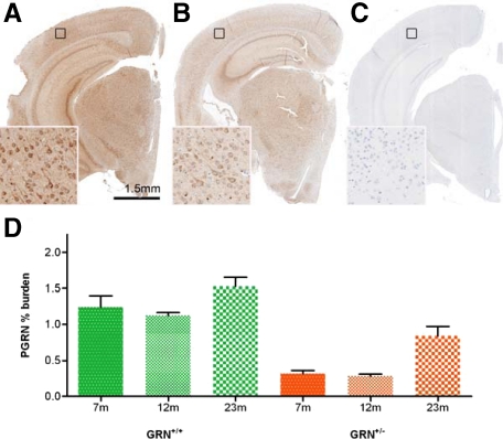
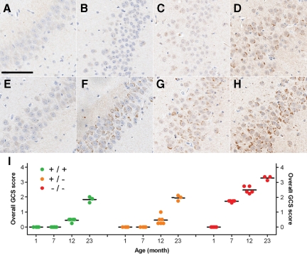
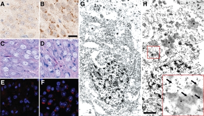
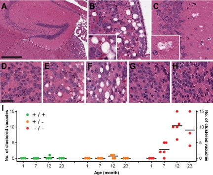
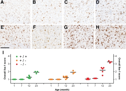
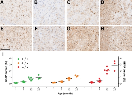
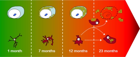
Progranulin (PGRN) is involved in wound repair, inflammation, and tumor formation, but its function in the central nervous system is unknown. Roles in development, sexual differentiation, and long-term neuronal survival have been suggested. Mutations in the GRN gene resulting in partial loss of the encoded PGRN protein cause frontotemporal lobar degeneration with ubiquitin immunoreactive inclusions. We sought to understand the neuropathological consequences of loss of PGRN function throughout the lifespan of GRN-deficient ((-/+) and (-/-)) mice. An aged series of GRN-deficient and wild-type mice were compared by histology, immunohistochemistry, and electron microscopy. Although GRN-deficient mice were viable, GRN(-/-) mice were produced at lower than predicted frequency. Neuropathologically, GRN(-/+) were indistinguishable from controls; however, GRN(-/-) mice developed age-associated, abnormal intraneuronal ubiquitin-positive autofluorescent lipofuscin. Lipofuscin was noted in aged GRN(+/+) mice at levels comparable with those of young GRN(-/-) mice. GRN(-/-) mice developed microgliosis, astrogliosis, and tissue vacuolation, with focal neuronal loss and severe gliosis apparent in the oldest GRN(-/-) mice. Although no overt frontotemporal lobar degeneration with ubiquitin immunoreactive inclusions type- or TAR DNA binding protein-43-positive lesions were observed, robust lipofuscinosis and ubiquitination in GRN(-/-) mice is strikingly similar to changes associated with aging and cellular decline in humans and animal models. Our data suggests that PGRN plays a key role in maintaining neuronal function during aging and supports the notion that PGRN is a trophic factor essential for long-term neuronal survival.


Figures

Figure 1
Figure 1
PGRN immunoreactivity is reflective of genotypes and increases in older GRN+/+ and GRN+/− mice. As shown in 12-month-old mice, GRN+/+ mice (A) have higher PGRN immunoreactivity than GRN+/− mice (B), whereas GRN−/− mice (C) have no detectable PGRN expression. Higher magnification insets correspond to the areas identified by black squares. D: In a subset of GRN+/+ (green) and GRN+/− (orange) mice, PGRN immunoreactivity was quantified by image analysis. Consistent with genotypes, GRN+/− mice had significantly reduced PGRN levels compared with GRN+/+ mice, at all ages (P < 0.05). An increase in PGRN immunoreactivity was also detected in 23-month-old GRN+/+ and GRN+/− mice compared with 7- and 12-month-old mice of the same genotype (P < 0.05). N = at least three for each group. Error bars = SEM. Scale bar = 1.5 mm.
Figure 2
Figure 2
Age-associated increase in ubiquitin-positive GCS. Ubiquitin immunohistochemistry demonstrating the age-associated increase in GCS in the CA2-3 region of the hippocampus of GRN+/+ (A–D) and GRN−/− (E–H) mice, with the latter showing an accelerated increase in GCS. A and E: 1 month. B and F: 7 months. C and G: 12 months. D and H: 23 months. GRN+/+ and GRN+/− showed no noticeable difference in GCS. I: A semiquantitative scoring scheme was used to measure the severity of GCS in key anatomical regions (Supplemental Figure 2, see http://ajp.amjpathol.org) to calculate an overall GCS score (mean of regional scores) for each mouse (colored circles). Overall GCS scores increased with age in all groups (P < 0.001). They also indicated that GCS occurred earlier (seven months) and was more severe in GRN−/− mice (P < 0.001 for 7, 12, and 23 months) compared with age-matched GRN+/+ and GRN+/− mice. The oldest GRN−/− mice had the highest overall GCS score. Horizontal black bars, mean. Scale bar = 100 μm.
Figure 3
Figure 3
Histological and ultrastructural properties of neuronal, ubiquitin-positive GCS. Ubiquitin immunohistochemistry identified GCS in neurons of the hippocampus (CA2-3 region) were largely absent from 23-month-old GRN+/+ (A; shown) and GRN+/− mice but were frequent in age-matched GRN−/− mice (B). Unlike the GRN+/+ mice (C and E), structures corresponding to GCS in GRN−/− mice were also highlighted by LFB-PAS staining (D) and were autofluorescent (F) as shown by 4′,6-diamidino-2-phenylindole staining viewed under a fluorescent microscope. These histological features are consistent with characteristics of lipofuscin. Ubiquitin-specific immunoelectron microscopy in 7-month-old mice confirmed the presence of lipofuscin granules (asterisk), which were qualitatively less numerous in CA2-3 neurons of GRN+/+ (G; shown) and GRN+/− mice compared with GRN−/− mice (H). Higher magnification inset shows gold particles (arrows) decorating clusters of granules containing a heterogeneous mixture of electron-dense material, consistent with ultrastructural properties of lipofuscin. N, nucleus. Scale bar: 25 μm A–F; 1 μm G and H.
Figure 4
Figure 4
Abnormal tissue vacuolation in older GRN−/− mice. H&E staining identified vacuolation in the habenular nucleus (A and B) and the neuropil surrounding CA2-3 regions (A and C) of the hippocampus in older GRN−/− mice. The former was largely characterized by single vacuoles whereas the latter contained mainly clustered vacuoles (inset). In GRN−/− mice, habenular vacuolation was not present at 1 month (D) but was a consistent feature in older GRN−/− (E: 7 month; B: 12 month; F: 23 month] mice and absent in all GRN+/+ (G) and GRN+/− mice (H), even at 23 months. I: Clustered vacuoles were counted in CA2-3 of the hippocampus, occurring in the majority of GRN−/− mice at seven months and becoming more frequent in 12- and 23-month-old mice. These vacuoles were largely absent in GRN+/+ and GRN+/−, but if present had a low frequency. Differences in the number of clustered vacuoles between GRN−/− mice and GRN+/+ or GRN+/− mice were statistically significant for 7-, 12-, and 23-month-old mice (P < 0.05). Horizontal black bars, mean. Scale bar: 400 μm A; 30 -μm D–H.
Figure 5
Figure 5
Changes in microglial morphology indicate increased microgliosis in GRN−/− mice. Regional examination of Iba-1-positive microglia in a 23-month-old GRN+/+ mouse showed a mixture of ramified and reactive microglia in the hippocampus CA2-3 region (A) and cortex (B) with predominately reactive microglia in the thalamus (C) and brainstem (D). In contrast, age-matched GRN−/− mice contained mainly amoeboid microglia in the hippocampus (E), reactive microglia in the cortex (F), and phagocytic microglia in the thalamus (G) and brainstem (H). GRN+/− mice (not shown) were indistinguishable from age-matched GRN+/+ mice. I: The morphology of Iba-1-positive microglia was scored in key anatomical regions (Supplemental Figure 3, see http://ajp.amjpathol.org) and averaged to generate an overall Iba-1 score for each mouse (individual circles). Overall Iba-1 scores were comparable across genotypes at one month of age; however, at 7, 12 and 23 months of age, GRN−/− mice had significantly higher scores (P < 0.001) compared with age-matched GRN+/+ and GRN+/− mice. Overall Iba-1 scores for 23-month-old GRN+/+ and GRN+/− mice was comparable with that of 12-month-old GRN−/− mice, whereas the oldest GRN−/− mice (23 months) had the highest overall Iba-1 score. Overall Iba-1 scores increased with age in all genotypic groups (P < 0.001). Horizontal black bars, mean; Iba-1 scores: 1, ramified; 2, reactive; 3, amoeboid; 4, phagocytic. Scale bar = 150 μm.
Figure 6
Figure 6
Increased astrogliosis in GRN−/− mice. The GFAP burden was comparatively lower in 23-month GRN+/+ and GRN+/− (not shown) mice in the CA2-3 (A), cortex (B), thalamus (C), and brainstem (D) compared with age-matched GRN−/− mice (E–H). The GFAP burden in GRN−/− mice was greatest in the thalamus (G) and brainstem (H), with CA2-3 (E) and cortex (F) having a lower burden. In these same regions, the degree of astrogliosis was quantified by measuring GFAP immunoreactivity (% burden) by image analysis for each mouse (individual circles, I). In younger mice (1–7 months), the mean GFAP burden (horizontal black bar) was similar among genotypic groups. Older GRN−/− mice (12 and 23 months) had a significantly higher GFAP burden (P < 0.01) compared with age-matched GRN+/+ and GRN+/− mice. The GFAP burden increased with age in all genotypic groups. Scale bar = 150 μm.
Figure 7
Figure 7
Hypothetical model of neurodegeneration based on the chronological appearance of pathology in GRN−/− mice. 1 month: young neurons are free of any pathology and microglia are in a ramified or resting state; seven months: through an unknown mechanism, the lack of PGRN results in intraneuronal accumulation of lipofuscin pigment in aging neurons resulting in cellular stress which is detected by nearby microglia; 12 months: neuronal cytoplasm is full of lipofuscin pigment which adversely affects cellular function and long-term neuronal survival, signaling the activation of microglia; 23 months: slow and progressive loss of neurons, resulting in expulsion of neuronal lipofuscin into extracellular space, most of which is phagocytosed by activated microglia with some lipofuscin accumulating in the extracellular space. The lack of PGRN may also affect the normal function of activated microglia resulting in accumulation of endogenous microglial lipofuscin. A severe, sustained, and/or an inappropriate microglia (inflammatory) response (attributable to absence of microglial PGRN) may further enhance neuronal loss.

Similar articles

Cited by

References

    1. Daniel R, Daniels E, He Z, Bateman A. Progranulin (acrogranin/PC cell-derived growth factor/granulin-epithelin precursor) is expressed in the placenta, epidermis, microvasculature, and brain during murine development. Dev Dyn. 2003;227:593–599. - PubMed
    1. Avrova AO, Stewart HE, De Jong WD, Heilbronn J, Lyon GD, Birch PR. A cysteine protease gene is expressed early in resistant potato interactions with Phytophthora infestans. Mol Plant Microbe Interact. 1999;12:1114–1119. - PubMed
    1. Bhandari V, Palfree RG, Bateman A. Isolation and sequence of the granulin precursor cDNA from human bone marrow reveals tandem cysteine-rich granulin domains. Proc Natl Acad Sci USA. 1992;89:1715–1719. - PMC - PubMed
    1. Ahmed Z, Mackenzie IR, Hutton ML, Dickson DW. Progranulin in frontotemporal lobar degeneration and neuroinflammation. J Neuroinflammation. 2007;4:7. - PMC - PubMed
    1. He Z, Bateman A. Progranulin (granulin-epithelin precursor, PC-cell-derived growth factor, acrogranin) mediates tissue repair and tumorigenesis. J Mol Med. 2003;81:600–612. - PubMed

Publication types