. 2010 Sep 9;116(10):1678-84.
doi: 10.1182/blood-2010-03-273862. Epub 2010 Jun 3.

A mutant allele of the Swi/Snf member BAF250a determines the pool size of fetal liver hemopoietic stem cell populations

Affiliations

A mutant allele of the Swi/Snf member BAF250a determines the pool size of fetal liver hemopoietic stem cell populations

Jana Krosl et al. Blood. .

Abstract

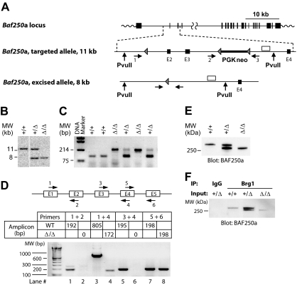
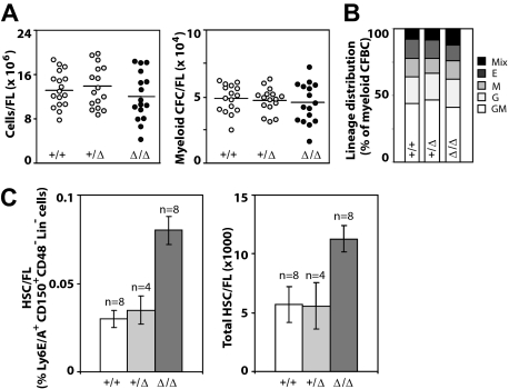
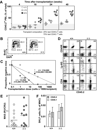
It is believed that hemopoietic stem cells (HSC), which colonize the fetal liver (FL) rapidly, expand to establish a supply of HSCs adequate for maintenance of hemopoiesis throughout life. Accordingly, FL HSCs are actively cycling as opposed to their predominantly quiescent bone marrow counterparts, suggesting that the FL microenvironment provides unique signals that support HSC proliferation and self-renewal. We now report the generation and characterization of mice with a mutant allele of Baf250a lacking exons 2 and 3. Baf250a(E2E3/E2E3) mice are viable until E19.5, but do not survive beyond birth. Most interestingly, FL HSC numbers are markedly higher in these mice than in control littermates, thus raising the possibility that Baf250a determines the HSC pool size in vivo. Limit dilution experiments indicate that the activity of Baf250a(E2E3/E2E3) HSC is equivalent to that of the wild-type counterparts. The Baf250a(E2E3/E2E3) FL-derived stroma, in contrast, exhibits a hemopoiesis-supporting potential superior to the developmentally matched controls. To our knowledge, this demonstration is the first that a mechanism operating in a cell nonautonomous manner canexpand the pool size of the fetal HSC populations.


Figures

Figure 1
Figure 1
Generation of Baf250aE2E3/E2E3 mouse strain. (A) Baf250a targeting strategy. Top, Baf250a locus (NM_001080819_MGI:1935147_Arid1a). Middle, the targeted allele before Cre-mediated excision. Bottom, the recombined Baf250aE2E3 allele. Exons (■), LoxP sites (▴), primers for PCR-based genotyping (numbered horizontal arrows), and 3′ external genomic probe (□) are shown. Positions of PvuII restriction sites are also denoted. (B) Southern blot analysis of Baf250a modification. DNA isolated from embryonic tissues was digested with PvuII, which cuts outside of the targeted region to generate a 11.6 kb WT and a 7.9 kb Baf250aE2E3 bands recognized by the 3′ probe depicted in Figure 1A. (C) PCR-based amplifications of Baf250aE2E3 allele. The primer pair 1 + 3 generates a 210-bp fragment identifying the excised Baf250aE2E3, and primers 2 + 3 pair generate the 90-bp WT fragment. Positions of primers are denoted in Figure 1A. (D) Quantitative RT-PCR–based analysis of Baf250aE2E3 mRNA. Note the absence of RT-PCR signal for homozygous Baf250aE2E3 cells when using primers positioned in exons 2 or 3, and the 172-bp segment amplified with primers in exons 1 and 4. Top, schema of primer positions. Bottom, amplicons generated by the denoted primer pairs. (E) Western blot analysis of BAF250a levels in WT, Baf250aE2E3, and Baf250aE2E3/E2E3 embryonic fibroblasts. The BAF250a antibody recognizes the approximately 280 kDa WT, and approximately 250 kDa mutated BAF250a proteins. (F) BAF250aE2E3 protein interacts with Brg1. Anti-Brg1 antibody coimmunoprecipitates WT and mutated BAF250a.
Figure 2
Figure 2
Hemopoietic characterization of E14.5 dpc Baf250aE2E3/E2E3 FL cell populations. (A) Total number of FL cells (left) and number of myeloid clonogenic progenitors (right) in FL. (B) Frequency of distribution of different myeloid progenitors in FL. (C) The frequency of HSCs in E14.5 FL (left) as defined by the CD150+Ly6A/E+CD48Lin phenotype. The total number of HSCs in E14.5 FL (right). The HSC numbers were calculated from total mononuclear cell numbers and the HSC frequency determined for each FL population. Shown are mean (± SD). Genotypes of the examined FL populations are shown at with image. Mix indicates CFU-granulocyte, erythrocyte, monocyte/macrophage, megakaryocyte; E, CFU-erythroid; M, CFU-monocyte/macrophage; G, CFU-granulocyte; and GM, CFU-granulocyte-monocyte/macrophage.
Figure 3
Figure 3
Effect of Baf250aE2E3 on HSC activity. (A) Homozygosity at Baf250aE2E3 allele enhances the competitive repopulation ability of mutant FL cells. WT, Baf250aE2E3, or Baf250aE2E3/E2E3 (CD45.2+) FL cells were mixed in a 1:4 ratio with CD45.1+ WT FL cells, and transplanted into lethally irradiated CD45.1+ recipient mice. Contribution of donor cells in peripheral blood of recipient mice was assessed at different times after transplantation. (B) BrdU incorporation profile of the CD150+CD48Lin cell population was assessed after 30 minutes in vivo BrdU pulse. Cell numbers are mean (± SD) values obtained from 3 developmentally matched FL. (C) Limiting dilution analysis for estimation of CRU frequency in WT and mutant FL (2 FL populations and 2 cohorts of recipient mice for each genotype). (D) Lympho-myeloid repopulation of recipient mice transplanted 20 weeks before with 100 000 WT or 10 000 Baf250aE2E3/E2E3 FL cells. Representative FACS profiles demonstrate comparable contributions of the transplant cell populations to repopulation of myeloid (Ly6G+), B lymphoid (CD45R+), and T lymphoid (CD4+) PBL. Average value in each quadrant represents number determined for mice (n = 5 in each group). (E) Proliferation potential of the transplanted HSCs. MAS, or proportion of PBL generated by individual transplant test CRU, was determined for groups of mice (n = 12) transplanted with low numbers (1-3) of Ly45.2+ WT or Baf250aE2E3/E2E3 FL CRU, together with 10 BM-derived competitor Ly45.1+CRU. (F) Contributions of the transplanted WT and Baf250aE2E3/E2E3 CRUs to regeneration of adult HSC pool. Proportions of test CD45.2+ and competitor CD45.1+-derived CD117+Ly6A/E+ (CD135Lin cells) in the BM of recipients described for Figure 1E were determined at 24 weeks after transplantation. Results represent mean (± SD) determined for each group of recipients (n = 5).
Figure 4
Figure 4
Baf250aE2E3/E2E3 stromal cell cultures support the in vitro self-renewal of progenitor/stem cell populations. (A) Enumeration of mesenchymal stem/progenitor cells in WT, Baf250aE2E3/+, and Baf250aE2E3/E2E3 FL cell populations. Each circular symbol represents individual FL population. Note the statistically significant difference between the WT, or Baf250aE2E3/+, and the Baf250aE2E3/E2E3 FL (unpaired t test, and 2-tailed P value determined). (B) Maintenance of CAFCday28 in the presence of WT or Baf250aE2E3/E2E3 stromal cells. Primary stromal cell cultures (4-cm - 10-cm dishes for each genotype) were established using pools of 4 FL cell populations. BM cells isolated from recipients of WT or Baf250aE2E3/E2E3 FL (2 recipients per genotype, > 85% of donor-derived PBL) were cocultured with stromal cell cultures for 28 days. CAFCday28 frequency at the beginning of experiment (t0), or after 28-day maintenance in stromal cocultures, were determined by plating an aliquot of the input BM cells, or entire cellular content of the stromal cocultures, at limit dilution on pre-established AFT024 cell layers. Results are mean (± SD), of 2 independent experiments performed in duplicate and are presented as the number of CAFC per 3 × 10⁷ input BM cells. (C) Baf250aE2E3/E2E3 stromal cell cultures support self-renewal of progenitor/stem cell populations. WT BM cells were seeded at limit dilution on pre-established WT or Baf250aE2E3/E2E3 stromal cell layers in 96-well trays (4 independent stromal cell cultures for each genotype). After 4-week coculture, all wells estimated to comprise a single cobblestone area were replated on freshly established WT or Baf250aE2E3/E2E3 stromal cell layers, and the frequency of secondary CAFCday28 was determined after additional 4-week maintenance.

References

    1. Mikkola HK, Orkin SH. The journey of developing hematopoietic stem cells. Development. 2006;133(19):3733–3744. - PubMed
    1. Ema H, Nakauchi H. Expansion of hematopoietic stem cells in the developing liver of a mouse embryo. Blood. 2000;95(7):2284–2288. - PubMed
    1. Bowie MB, McKnight KD, Kent DG, McCaffrey L, Hoodless PA, Eaves CJ. Hematopoietic stem cells proliferate until after birth and show a reversible phase-specific engraftment defect. J Clin Invest. 2006;116(10):2808–2816. - PMC - PubMed
    1. Foudi A, Hochedlinger K, Van BD, et al. Analysis of histone 2B-GFP retention reveals slowly cycling hematopoietic stem cells. Nat Biotechnol. 2009;27(1):84–90. - PMC - PubMed
    1. Wilson A, Laurenti E, Oser G, et al. Hematopoietic stem cells reversibly switch from dormancy to self-renewal during homeostasis and repair. Cell. 2008;135(6):1118–1129. - PubMed

Publication types

MeSH terms