. 2010 May 27;5(5):e10859.
doi: 10.1371/journal.pone.0010859.

miR-22 forms a regulatory loop in PTEN/AKT pathway and modulates signaling kinetics

Affiliations

miR-22 forms a regulatory loop in PTEN/AKT pathway and modulates signaling kinetics

Nadav Bar et al. PLoS One. .

Abstract

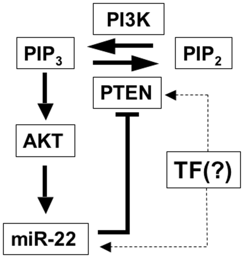
Background: The tumor suppressor PTEN (phosphatase and tensin homolog) is a lipid phosphatase that converts PIP3 into PIP2 and downregulates the kinase AKT and its proliferative and anti-apoptotic activities. The FoxO transcription factors are PTEN downstream effectors whose activity is negatively regulated by AKT-mediated phosphorylation. PTEN activity is frequently lost in many types of cancer, leading to increased cell survival and cell cycle progression.

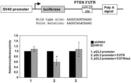
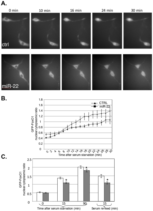
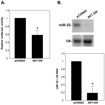
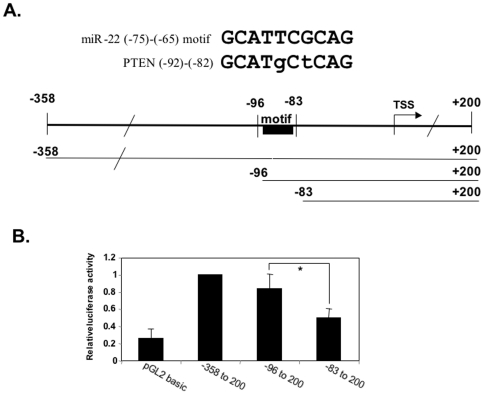
Principal findings: Here we characterize the widely expressed miR-22 and report that miR-22 is a novel regulatory molecule in the PTEN/AKT pathway. miR-22 downregulates PTEN levels acting directly through a specific site on PTEN 3'UTR. Interestingly, miR-22 itself is upregulated by AKT, suggesting that miR-22 forms a feed-forward circuit in this pathway. Time-resolved live imaging of AKT-dependent FoxO1 phosphorylation revealed that miR-22 accelerated AKT activity upon growth factor stimulation, and attenuated its down regulation by serum withdrawal.

Conclusions: Our results suggest that miR-22 acts to fine-tune the dynamics of PTEN/AKT/FoxO1 pathway.


Conflict of interest statement

Competing Interests: The authors have declared that no competing interests exist.

Figures

Figure 1
Figure 1. Identification of miR-22 promoter.
A. A 1155 bp fragment of miR-22 predicted promoter from −1100 to +55 was cloned upstream to the luciferase gene in the promoter-less pGL2 plasmid and transfected into HEK 293T together with RSV-renilla that served as internal control for transfection efficiency. pGL2-basic and pGL2-SV40-promoter were also transfected and served as negative and positive controls respectively. 24 hours later the relative luciferase activities were determined. The results represent the mean +/− SD of 7 independent transfection experiments. B. Determination of miR-22 TSS. The luciferase reporter gene under the control of miR-22 promoter was transfected into HEK293T cells and 24 hours after transfection RNA was extracted and analyzed by primer extension using a primer complementary to the 5′-end of the luciferase gene. The labeled miR-22 derived cDNA was run on a denaturing 8% urea-polyacrylamid gel alongside a sequencing reaction marked by A, G, C and T. C. Analysis of sensitivity of endogenous miR-22 transcription to α-amanitin. Nuclei were isolated from HEK293T cells and subjected to a run-on assay in the absence or presence of 20 µg/ml α-amanitin using 32P-labeled UTP. Labeled RNAs were isolated and then hybridized to membranes that were dot-blotted with RNA pol I gene, 45s rRNA; RNA pol III gene tRNAY and miR-22 transcripts as indicated. D. Analysis of miR-22 promoter organization. The miR-22 promoter was dissected from the 5′ as shown schematically and luciferase activity of the different constructs was determined as described in A. The results represent the mean +/− SD of 4–6 independent transfection experiments.
Figure 2
Figure 2. PTEN is a direct target of miR-22.
A. Northern blot analysis with miR-22 and U6 probes of small RNAs extracted from HeLa or HEK293T cells, as indicated, transfected with miR-22 or parental (pCDNA3) expression plasmids. B. Left panel shows western blot analyses, with PTEN, tubulin and GAPDH antibodies, of cell lystates prepared from HeLa, HEK293T and MCF-7 cells, as indicated, transfected with miR-22 expression plasmid or the parental vector pCDNA3. Quantification, by densitometry, of 3 independent transfection experiments in HeLa cells is shown in the right panel in which * indicates p<0.01. C. Representative western blot analysis with PTEN and GAPDH antibodies of cell lystates prepared from HeLa cells transfected with miR-22 or control antagomir expression plasmids. Quantification of PTEN levels of 4 independent transfection experiments is shown in the right panel in which * indicates p<0.05.
Figure 3
Figure 3. miR-22 targets PTEN through a site at the 3′UTR.
A schematic representation of a reporter gene used to analyze the miR-22 target site of PTEN 3′UTR is shown in the upper panel. The sequences of the wild type and mutated target site are shown. The wild type and mutated reporter plasmids were co-transfected into HEK293T cells together with miR-22 expression plasmid or the parental vector pCDNA3 and RSV-renilla that served as internal control. The activity of each construct without miR-22 was set to 1. The results represent the average +/− SD of four independent transfection experiments in which the bar marked with * indicates p<0.01.
Figure 4
Figure 4. The effect of miR-22 expression on the sub-cellular localization of FoxO1.
A. NIH3T3 cells were co-transfected with FoxO1-GFP together with either miR-22 expression plasmid or the parental pCDNA3. 48 hours after transfection FoxO1-GFP from 10 fields of each transfection was visualized by Time-Lapse microscopy before and after serum deprivation. Representative images from the indicated time points after serum withdrawal are shown. B. Quantitative analysis of changes in FoxO1-GFP localization after serum starvation at 2 minutes intervals up to 30 minutes. The measurement was carried out by densitometry of the fluorescent signal in the cytoplasm and the nucleus of each cell. The data is presented as the ratio between nucleus and cytoplasm. C. Analysis of changes in FoxO1-GFP subcellular localization as in B using fluorescent microscope. Images of 15–20 fields (30–50 cells) of each transfection were taken at four different time points: before starvation (time 0), 15 and 90 minutes after replacing the cell medium to serum-free medium, and 15 minutes after addition of serum to the sarved cells (Re-Feed). The results are the sum of three independent biological repeats. The asterisks indicate that the differences between control and miR-22 in 15, 90 minutes after starvation and reefed points are statistically significant (p = 8×10⁻⁵, 3×10⁻², and 2×10⁻⁴ respectively).
Figure 5
Figure 5. The PI3K/AKT pathway affects miR-22 expression.
A. The luciferase reporter gene under the control of miR-22 promoter was co-transfected into HEK293T cells together with either an empty expression plasmid (pCNDA3) or plasmid expressing a dominant negative mutant of AKT (AKT-DN). 24 hours after transfection cells were harvested and the relative luciferase activity was determined. The results represent the mean +/− SD of three independent experiments. The asterisk indicates statistically significant difference (p<0.01). B. HeLa cells were transfected with either dominant negative AKT or empty expression plasmid together with puromycin resistant gene plasmid. 24–48 hours after transfection small RNAs were extracted and subjected to Northern blot with miR-22 and U6 probes as indicated. A representative Northern blot is shown in the top panel and densitometric quantification of three independent transfection experiments is shown in the bottom panel. The asterisk indicates statistically significant difference (p<0.001).
Figure 6
Figure 6. Identification of an important transcription regulatory element in PTEN promoter that is similar to the −75 to −65 motif of miR-22 promoter.
The upper panel shows sequence alignment between the −75 to −65 motif of miR-22 promoter and the −92 to −82 region of PTEN promoter. Fragments of PTEN promoter with progressive 5′ deletions that are schematically shown, were cloned upstream to a luciferase gene in the promoter-less pGL2 basic and then transfected into HEK293T cells together with RSV-renilla that served as internal control for transfection efficiency. 24 hours later the relative luciferase activities were determined. The results represent the mean +/− SD of 7 independent transfection experiments. The bars marked with asterisk indicate statistically significant difference p<0.001.
Figure 7
Figure 7. A model showing PTEN/PI3K/AKT pathway with miR-22 and the regulatory loop exerted by miR-22.

References

    1. Cantley LC, Neel BG. New insights into tumor suppression: PTEN suppresses tumor formation by restraining the phosphoinositide 3-kinase/AKT pathway. Proc Natl Acad Sci U S A. 1999;96:4240–4245. - PMC - PubMed
    1. Di Cristofano A, Pandolfi PP. The multiple roles of PTEN in tumor suppression. Cell. 2000;100:387–390. - PubMed
    1. Greer EL, Brunet A. FOXO transcription factors at the interface between longevity and tumor suppression. Oncogene. 2005;24:7410–7425. - PubMed
    1. Huse JT, Brennan C, Hambardzumyan D, Wee B, Pena J, et al. The PTEN-regulating microRNA miR-26a is amplified in high-grade glioma and facilitates gliomagenesis in vivo. Genes Dev. 2009;23:1327–1337. - PMC - PubMed
    1. Kato M, Putta S, Wang M, Yuan H, Lanting L, et al. TGF-beta activates Akt kinase through a microRNA-dependent amplifying circuit targeting PTEN. Nat Cell Biol. 2009;11:881–889. - PMC - PubMed

Publication types

MeSH terms