. 2010 May 27;5(5):e10860.
doi: 10.1371/journal.pone.0010860.

Mycobacterium tuberculosis transcriptional adaptation, growth arrest and dormancy phenotype development is triggered by vitamin C

Affiliations

Mycobacterium tuberculosis transcriptional adaptation, growth arrest and dormancy phenotype development is triggered by vitamin C

Neetu Kumra Taneja et al. PLoS One. .

Abstract

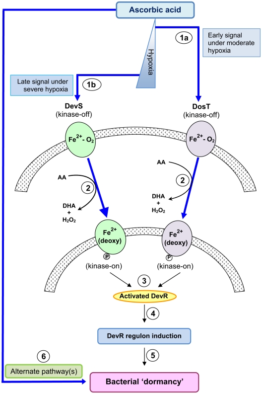
Background: Tubercle bacilli are thought to persist in a dormant state during latent tuberculosis (TB) infection. Although little is known about the host factors that induce and maintain Mycobacterium tuberculosis (M. tb) within latent lesions, O(2) depletion, nutrient limitation and acidification are some of the stresses implicated in bacterial dormancy development/growth arrest. Adaptation to hypoxia and exposure to NO/CO is implemented through the DevRS/DosT two-component system which induces the dormancy regulon.

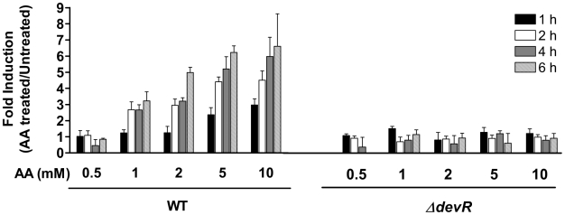
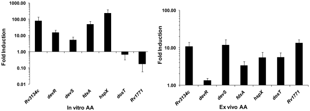
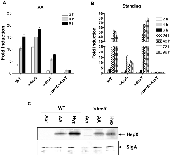
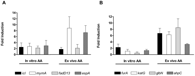
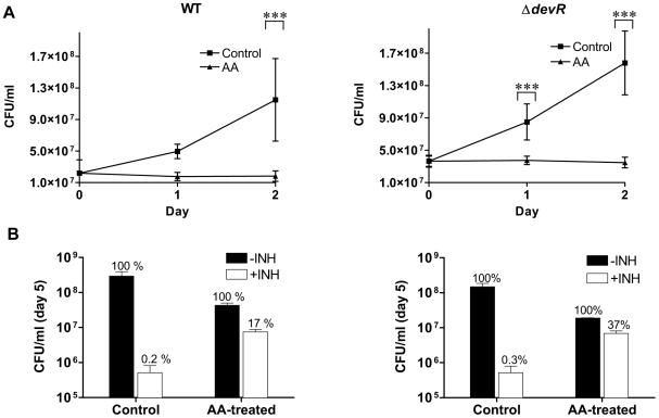
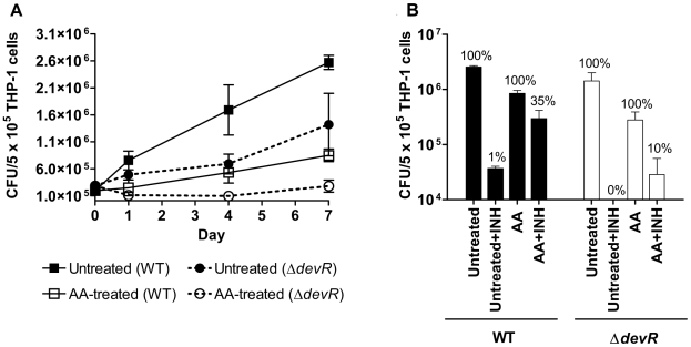
Methodology/principal findings: Here we show that vitamin C (ascorbic acid/AA) can serve as an additional signal to induce the DevR regulon. Physiological levels of AA scavenge O(2) and rapidly induce the DevR regulon at an estimated O(2) saturation of <30%. The kinetics and magnitude of the response suggests an initial involvement of DosT and a sustained DevS-mediated response during bacterial adaptation to increasing hypoxia. In addition to inducing DevR regulon mechanisms, vitamin C induces the expression of selected genes previously shown to be responsive to low pH and oxidative stress, triggers bacterial growth arrest and promotes dormancy phenotype development in M. tb grown in axenic culture and intracellularly in THP-1 cells.

Conclusions/significance: Vitamin C mimics multiple intracellular stresses and has wide-ranging regulatory effects on gene expression and physiology of M. tb which leads to growth arrest and a 'dormant' drug-tolerant phenotype, but in a manner independent of the DevRS/DosT system. The 'AA-dormancy infection model' offers a potential alternative to other models of non-replicating persistence of M. tb and may be useful for investigating host-'dormant' M. tb interactions. Our findings offer a new perspective on the role of nutritional factors in TB and suggest a possible role for vitamin C in TB.


Conflict of interest statement

Competing Interests: The authors have declared that no competing interests exist.

Figures

Figure 1
Figure 1. Induction of Rv3134c promoter activity in M. tuberculosis.
The induction of the Rv3134c promoter (in plasmid p3134c-1) was assessed by measuring GFP fluorescence in 0–6 hours standing M. tb cultures. Fold induction of promoter activity in AA-exposed cultures is expressed with respect to untreated cultures, both under standing conditions. Results are expressed as mean ± SD of 3 independent experiments.
Figure 2
Figure 2. AA-mediated induction of M. tb DevR regulon genes.
Real time RT-PCR analysis of selected DevR regulon genes was carried out with RNA isolated from M. tb H37Rv cultures treated in vitro with AA and M. tb RNA isolated from infected THP-1 cells that were treated with AA post-infection (8 hour exposure to AA for both). ‘In vitro AA’, fold induction of individual gene transcription is a ratio of 16S rRNA-normalized transcript number in AA-treated vs. untreated control cultures. Results are expressed as mean ± SD of 3 independent cultures each analyzed in 2-4 replicate assays. ‘Ex vivo AA’, fold induction of individual gene transcription is a ratio of 16S rRNA-normalized transcript number in AA-treated infected THP-1 cells vs. untreated infected THP-1 cells. Results are expressed as mean ± SD of pools of 10 independent infections, each pool analyzed in triplicate.
Figure 3
Figure 3. AA-mediated O2 scavenging results in induction of DevR-regulated promoter activity.
(A) Time dependent depletion of dissolved O2 (DO) in Dubos medium with AA concentrations of 1 mM (Δ), 2 mM (○), 5 mM (•), 10 mM (□), 10 mM DHA (×), expressed as% of initial value (saturation). (B) Rv1738 promoter activity leads to induction of GFP fluorescence (expressed in RFU/OD) in AA-treated shaking M. tb cultures. Data is shown as mean ± SD (n = 6) for 0 mM (▪), 1 mM (Δ), 2 mM (○), 5 mM (•), 10 mM (□) of AA. Inset in (B) shows promoter activity in AA-treated cultures at 24 h. The first time point showing a statistically significant induction is shown by the red arrow on the red line in (A). The data that was subjected to statistical analysis is shown in a blue shaded rectangle.
Figure 4
Figure 4. Differential signaling through DevS/DosT by AA and under gradual hypoxia.
Induction of Rv3134c promoter activity (GFP fluorescence in RFU/OD) was assessed in M. tb cultures treated with (A) 10 mM AA, or (B) standing for various intervals of time. Fold induction is the ratio between GFP fluorescence (in RFU/OD) in treated vs. untreated cultures, all in 96-well microplate standing format. (C) Western blot analysis of lysates for expression of HspX and SigA proteins in aerobic M. tb cultures (Aer) and those exposed for 8 h to 10 mM AA (under shaking aerobic conditions) or in 5-day standing (Hyp) cultures. Results are expressed as mean ± SD of 3 independent experiments. Note that the y-axis range is different in (A) and (B) panels.
Figure 5
Figure 5. AA-mediated induction of M. tb acid response and antioxidant response genes.
Experimental details are described in Fig. 2legend and Experimental procedures.
Figure 6
Figure 6. AA-triggered bacterial growth arrest and dormant physiology in axenic cultures of M. tb.
(A) Bacteriostasis of AA-treated aerobic M. tb cultures. Statistically significant difference in CFUs recovered from AA-treated vs. untreated (Control) aerobic cultures was determined using paired two-way ANOVA with Bonferroni post-test correction (***, p-value <0.001). (B) INH tolerance of AA-treated aerobic M. tb cultures. Cultures were exposed to 10 mM AA for 1 day followed by treatment with INH (4 µg/ml) for 4 days. The number of viable bacteria on day 5 is shown. CFU in ‘-INH’ ‘Control’ (untreated) or ‘AA-treated’ wells is depicted as 100%; bacteria surviving INH treatment are depicted as ‘+INH’ with respect to their controls (100%). Mean ± SD of 3 independent cultures is shown.
Figure 7
Figure 7. AA-triggered bacterial growth arrest and dormant physiology of M. tb in infected THP-1 cells.
(A) Bacteriostasis within untreated and AA-treated THP-1 cells. In case of ‘AA’-treated THP-1, the cells were treated with AA immediately after bacterial infection (i.e. day 0). Statistically significant difference in CFUs recovered from AA-treated vs. untreated THP-1 cells on various days was determined using paired two-way ANOVA with Bonferroni post-test correction (***, p-value <0.001). (B) Dormant physiology of M. tb within AA-treated THP-1 cells. Infected THP-1 cells were either not treated (‘Untreated’) or treated with 2 mM AA (‘AA’) for 7 days post-infection. INH (4 µg/ml final concentration) was added on day 1 to ‘Untreated+INH’ or ‘AA+INH’cultures, and INH treatment continued until day 7. Viable counts were determined on day 7 post-infection (i.e. 6 days post INH-treatment). ‘Untreated’, 100%, total bacteria on day 7; ‘Untreated+INH”, % bacterial recovery with reference to ‘Untreated’; AA, 100%, total bacteria on day 7; ‘AA+INH’, % bacterial recovery with reference to ‘AA’. Mean ± SD of 3 independent experiments is shown.
Figure 8
Figure 8. M. tb ‘dormancy’ response to ascorbic acid.
In one pathway, hypoxia generation by AA is sensed by DosT and DevS resulting in their activation (first deoxy-DosT and then deoxy-DevS under progressive hypoxia, steps 1a, 1b and 2). The transfer of phosphosignal from the sensors to DevR results in its activation (3). Activated DevR binds to target gene promoters and triggers DevR regulon mechanisms (4) and bacterial ‘dormancy’ (5). Bacterial ‘dormancy’ is also attained through alternate pathway(s) upon exposure to vitamin C (6).

Similar articles

Cited by

References

    1. Gomes MS, Paul S, Moreira AL, Appelberg R, Rabinovitch M, et al. Survival of Mycobacterium avium and Mycobacterium tuberculosis in acidified vacuoles of murine macrophages. Infect Immun. 1999;67:3199–3206. - PMC - PubMed
    1. Rohde KH, Abramovitch RB, Russell DG. Mycobacterium tuberculosis invasion of macrophages: linking bacterial gene expression to environmental cues. Cell Host Microbe. 2007;2:352–364. - PubMed
    1. Schnappinger D, Ehrt S, Voskuil MI, Liu Y, Mangan JA, et al. Transcriptional Adaptation of Mycobacterium tuberculosis within Macrophages: Insights into the Phagosomal Environment. J Exp Med. 2003;198:693–704. - PMC - PubMed
    1. Betts JC, Lukey PT, Robb LC, McAdam RA, Duncan K. Evaluation of a nutrient starvation model of Mycobacterium tuberculosis persistence by gene and protein expression profiling. Mol Microbiol. 2002;43:717–731. - PubMed
    1. Voskuil MI, Schnappinger D, Visconti KC, Harrell MI, Dolganov GM, et al. Inhibition of respiration by nitric oxide induces a Mycobacterium tuberculosis dormancy program. J Exp Med. 2003;198:705–713. - PMC - PubMed

Publication types

MeSH terms