. 2009 Dec 4;8(4):290-305.
doi: 10.2174/187152309789838975.

ANTI-INFECTIVE PROTECTIVE PROPERTIES OF S100 CALGRANULINS

Affiliations

ANTI-INFECTIVE PROTECTIVE PROPERTIES OF S100 CALGRANULINS

Kenneth Hsu et al. Antiinflamm Antiallergy Agents Med Chem. .

Abstract

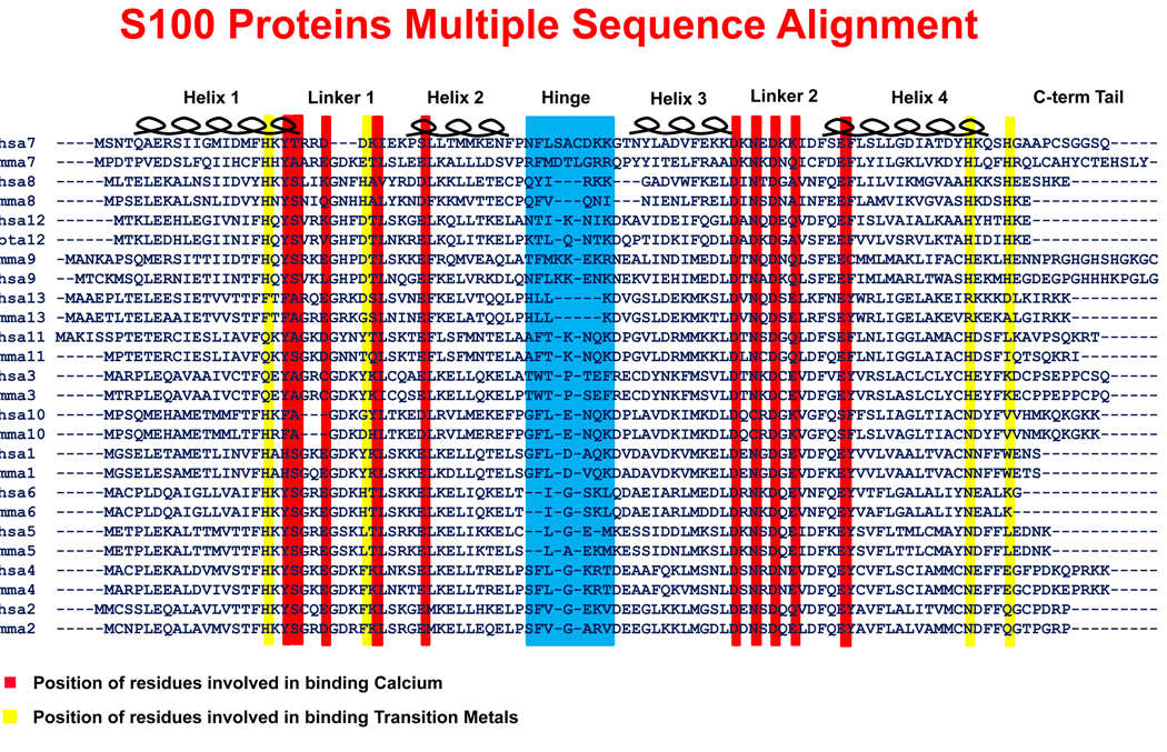
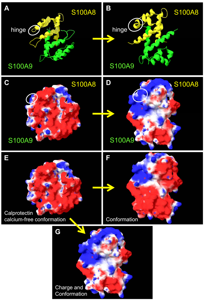
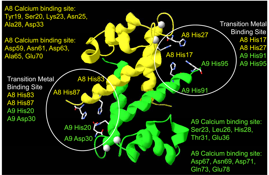
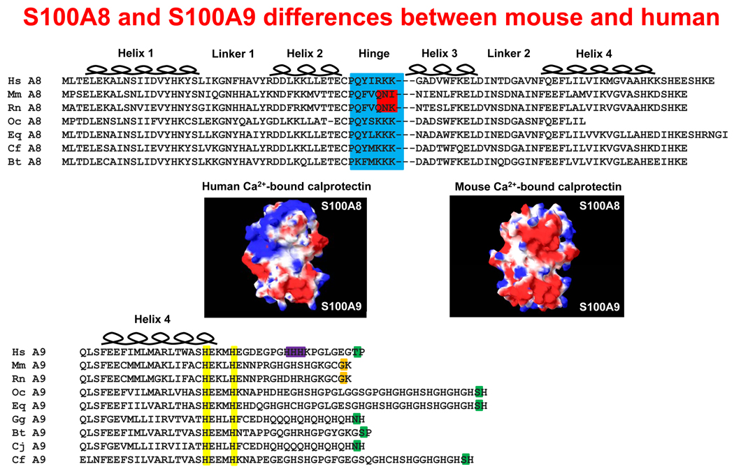
The calgranulins are a subgroup of proteins in the S100 family (calgranulin A, S100A8; calgranulin B, S100A9 and calgranulin C, S100A12) that provide protective anti-infective and anti-inflammatory functions for the mammalian host. In this review, we discuss the structure-function relationships whereby S100A8 and S100A9, and for comparison, S100A12, provide intra- and extracellular protection during the complex interplay between infection and inflammation and how the calgranulins are regulated to optimally protect the host. Ideally located to support epithelial barrier function, calprotectin, a complex of S100A8/S100A9, is expressed in squamous mucosal keratinocytes and innate immune cells present at mucosal surfaces. The calgranulins are also abundantly produced in neutrophils and monocytes, whereas expression is induced in epidermal keratinocytes, gastrointestinal epithelial cells and fibroblasts during inflammation. The calgranulins show species-specific expression and function. For example, S100A8 is chemotactic in rodents but not in humans. In humans, S100A12 appears to serve as a functional chemotactic homolog to murine S100A8. Transition metal-binding and oxidation sites within calgranulins are able to create structural changes that may orchestrate new protective functions or binding targets. The calgranulins thus appear to adopt a variety of roles to protect the host. In addition to serving as a leukocyte chemoattractant, protective functions include oxidant scavenging, antimicrobial activity, and chemokine-like activities. Each function may reflect the concentration of the calgranulin, post-transcriptional modifications, oligomeric forms, and the proximal intracellular or extracellular environments. Calprotectin and the calgranulins are remarkable as multifunctional proteins dedicated to protecting the intra- and extracellular environments during infection and inflammation.


Figures

Figure 1
Figure 1. Multiple sequence alignment of S100 protein family
A pair of sequences, mouse and human if available, for the S100A1 thru S100A13 proteins was used to generate the alignment. The common features of the S100 proteins are the first EF Hand (containing helices 1 & 2 as well as the linker 1 region), the hinge region, the second EF Hand (containing helices 3 & 4 as well as the linker 2 region), and the carboxy-terminal tail. A red background indicates the locations of the residues responsible for binding calcium. Although no S100 monomer is able to bind a transition metal, a pair of yellow lines indicates the location of the residues that create each half of the transition metal binding site within a dimer. The hinge region is highlighted in blue. The hinge region and helix 3 in S100A8 are shortened relative to other S100 proteins.
Figure 2
Figure 2. Predicted surface structures of S100A8 and S100A9 in the presence and absence of calcium
The ribbon diagrams (S100A8, yellow and S100A9, green) of (A) calcium-free and (B) calcium-bound human calprotectin. The white circle indicates the hinge region of S100A8 and highlights the random coil to alpha helix transition upon calcium-binding. The solvent accessible surfaces of (C) calcium-free and (D) calcium-bound calprotectin: blue represents positive potential, red is negative potential, and white indicates a hydrophobic region. (E) The conformation and charge of the solvent accessible surface of the calprotectin calcium-free model structure (as in C). (F) The changes in the molecular surface of calprotectin upon binding calcium without taking into account the positive charge associated with the calcium ion. This electrostatic component is added to the calculation for panel G. (G) Calcium-binding induces a conformation change and neutralizes a large area of negatively-charged surface of calprotectin.
Figure 3
Figure 3. Ribbon diagram of calcium-bound calprotectin showing likely transition metal binding sites
S100A8 (yellow ribbon) and S100A9 (green) residues coordinating calcium are listed, and coloured according to their location binding sites (highlighted with white circles) in S100A8 or S100A9. Coordination is created by the dimer interface and the ligands are coloured accordingly.
Figure 4
Figure 4. Structural comparision of murine and human calprotectin
Alignment of seven species of S100A8 sequences (top panel). The hinge region (highlighted in blue) contains three positively-charged residues, except in the case of mouse and rat, which have polar glutamine and asparagine residues (highlighted in red). In the calcium-bound conformation, the electrostatic potential differences for human and mouse calprotectin are given (middle panel). The solvent-accessible surfaces are represented: blue indicates positive potential, red shows negative potential, and white indicates a hydrophobic region. The multiple sequence alignment shows the penultimate C-terminal phosphorylatable residues (highlighted in green) and the non-phosphorylatable equivalent residues in murine and rat S100A9 (orange) (bottom panel). Residues involved in binding transition metals (yellow) and possible arachidonic acid binding (purple) are indicated.

References

    1. Foell D, Frosch M, Sorg C, Roth J. Phagocyte-specific calcium-binding S100 proteins as clinical laboratory markers of inflammation. Clin Chim Acta. 2004;344(1–2):37–51. - PubMed
    1. Salama I, Malone PS, Mihaimeed F, Jones JL. A review of the S100 proteins in cancer. Eur J Surg Oncol. 2008;34(4):357–364. - PubMed
    1. Berntzen HB, Fagerhol MK. L1, a major granulocyte protein: antigenic properties of its subunits. Scand J Clin Lab Invest. 1988;48(7):647–652. - PubMed
    1. Kerkhoff C, Klempt M, Sorg C. Novel insights into structure and function of MRP8 (S100A8) and MRP14 (S100A9) Biochim Biophys Acta. 1998;1448(2):200–211. - PubMed
    1. Honig B, Nicholls A. Classical electrostatics in biology and chemistry. Science. 1995;268(5214):1144–1149. - PubMed