

A case of leukemia cutis at the site of a prior catheter insertion
- PMID: 20523785
- PMCID: PMC2861221
- DOI: 10.5021/ad.2009.21.2.193
A case of leukemia cutis at the site of a prior catheter insertion
Abstract
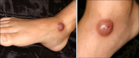
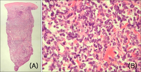
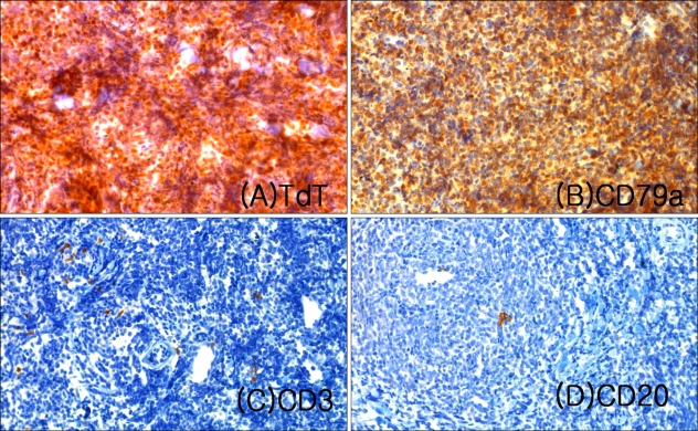
Leukemia cutis is the cutaneous involvement of leukemic neoplastic cells. It is an uncommon feature of systemic leukemia, and is associated with a poor prognosis. A 20-year-old man presented with a solitary, 2 cm domeshaped, firm, erythematous nodule on the right medial malleolus that was present for 3 months. The patient had a history of venous catheter insertion at the right medial malleolus area 3 months previously. The history was significant for acute lymphoblastic leukemia (ALL) for 4 years; an allogenic bone marrow transplantation was performed 3 years earlier. The histologc examination of the lesion revealed diffuse infiltration of leukemic cells in the dermis and subcutis. Herein we report a case of leukemia cutis at the site of a prior catheter insertion in a patient with ALL.
Keywords: Acute lymphocytic leukemia; Leukemia cutis; Site of catheter insertion.
Figures
Similar articles
-
Leukemia Cutis: An Unusual Presentation of Acute Lymphoblastic Leukemia in a Child.Indian J Dermatol. 2015 Nov-Dec;60(6):636. doi: 10.4103/0019-5154.169153. Indian J Dermatol. 2015. PMID: 26677299 Free PMC article.
-
Acral Solitary Nodule as a First Clinical Manifestation of Chronic Lymphocytic Leukemia.Adv Biomed Res. 2023 Aug 31;12:222. doi: 10.4103/abr.abr_268_22. eCollection 2023. Adv Biomed Res. 2023. PMID: 38073740 Free PMC article.
-
Aleukemic leukemia cutis manifesting with disseminated nodular eruptions and a plaque preceding acute monocytic leukemia: a case report.Case Rep Oncol. 2011 Sep;4(3):547-54. doi: 10.1159/000334745. Epub 2011 Nov 22. Case Rep Oncol. 2011. PMID: 22187541 Free PMC article.
-
Leukemia cutis presenting with fingertip hypertrophy.J Cutan Med Surg. 2003 Jan-Feb;7(1):57-60. doi: 10.1177/120347540300700110. Epub 2002 Oct 9. J Cutan Med Surg. 2003. PMID: 12362263 Review.
-
Leukemia cutis in congenital leukemia. Analysis and review of the world literature with report of an additional case.Arch Dermatol. 1993 Oct;129(10):1301-6. doi: 10.1001/archderm.129.10.1301. Arch Dermatol. 1993. PMID: 8215495 Review.
Cited by
-
Leukemia cutis as an initial presentation in a case of mixed phenotype acute leukemia: a double jeopardy.Int J Clin Exp Pathol. 2024 Aug 15;17(8):252-256. doi: 10.62347/ACDG7634. eCollection 2024. Int J Clin Exp Pathol. 2024. PMID: 39262435 Free PMC article.
-
Diagnostic Approaches in Myeloid Sarcoma.Curr Issues Mol Biol. 2025 Feb 10;47(2):111. doi: 10.3390/cimb47020111. Curr Issues Mol Biol. 2025. PMID: 39996833 Free PMC article. Review.
-
Leukemia Cutis: An Unusual Presentation of Acute Lymphoblastic Leukemia in a Child.Indian J Dermatol. 2015 Nov-Dec;60(6):636. doi: 10.4103/0019-5154.169153. Indian J Dermatol. 2015. PMID: 26677299 Free PMC article.
-
Leukaemia cutis for clinicians, a literature review.Postepy Dermatol Alergol. 2021 Jun;38(3):359-365. doi: 10.5114/ada.2021.107923. Epub 2021 Jul 26. Postepy Dermatol Alergol. 2021. PMID: 34377113 Free PMC article. Review.
-
[Clinical analysis of skin infiltration in acute leukemia diagnosed by fine needle aspiration cytology].Zhonghua Xue Ye Xue Za Zhi. 2021 Jan 14;42(1):78-80. doi: 10.3760/cma.j.issn.0253-2727.2021.01.015. Zhonghua Xue Ye Xue Za Zhi. 2021. PMID: 33677874 Free PMC article. Chinese. No abstract available.
References
-
- Su WP, Buechner SA, Li CY. Clinicopathologic correlations in leukemia cutis. J Am Acad Dermatol. 1984;11:121–128. - PubMed
-
- Koizumi H, Kumakiri M, Ishizuka M, Ohkawara A, Okabe S. Leukemia cutis in acute myelomonocytic leukemia: infiltration to minor traumas and scars. J Dermatol. 1991;18:281–285. - PubMed
-
- Baden TJ, Gammon WR. Leukemia cutis in acute myelomonocytic leukemia. Preferential localization in a recent Hickman catheter scar. Arch Dermatol. 1987;123:88–90. - PubMed
-
- Burns CA, Scott GA, Miller CC. Leukemia cutis at the site of trauma in a patient with Burkitt leukemia. Cutis. 2005;75:54–56. - PubMed
-
- Kim JE, Kim MY, Kim HO, Park YM. Plasma cell leukaemia cutis preferentially localized to recent puncture sites. Br J Dermatol. 2004;151:237–238. - PubMed
Publication types
LinkOut - more resources
Full Text Sources
