

A Case of Pigmented Bowen's Disease
- PMID: 20523786
- PMCID: PMC2861209
- DOI: 10.5021/ad.2009.21.2.197
A Case of Pigmented Bowen's Disease
Abstract
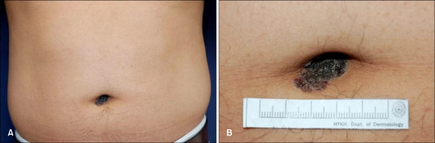
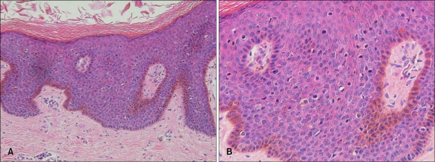
Pigmented Bowen's disease is characterized by increased melanin pigment in the epidermis or papillary dermis, in addition to the typical findings of Bowen's disease. This disorder has been infrequently reported and it represents less than 2% of all cases of Bowen's disease. Thus far, there has been only 1 case of pigmented Bowen's disease on the umbilicus in the medical literature, and no such case has been reported in Korea. Pigmented Bowen's disease develops on sun-exposed areas of the face and neck, as well as sun-unexposed areas like the trunk, extremities, perianal area and umbilcus. A 36-year-old man presented with a 9-month history of solitary dark brown slithery or wavy plaque with a verrucous surface on the umbilicus, and the lesion measured 1 x 2 cm in size. The histopathologic findings showed hyperkeratosis, parakeratosis and atypical keratinocytes disorderly arranged throughout the epidermis. Increased melanin pigment was noted in the basal layer of the epidermis. From these findings, we diagnosed this lesion as pigmented Bowen's disease.
Keywords: Bowen's disease; Pigmented; Umbilicus.
Figures
Similar articles
-
Pigmented lesion in the inguinal region.Dermatol Online J. 2011 Nov 15;17(11):12. Dermatol Online J. 2011. PMID: 22136868
-
Reflectance Confocal Microscopy of Pigmented Bowen's Disease: A Case Series of Difficult to Diagnose Lesions.Case Rep Dermatol. 2020 May 15;12(2):98-106. doi: 10.1159/000507916. eCollection 2020 May-Aug. Case Rep Dermatol. 2020. PMID: 32518541 Free PMC article.
-
Pigmented Bowen's Disease: A Case Report in Saudi Arabia.Cureus. 2024 Aug 5;16(8):e66191. doi: 10.7759/cureus.66191. eCollection 2024 Aug. Cureus. 2024. PMID: 39233960 Free PMC article.
-
Pigmented Bowen's disease and review of 420 Bowen's disease lesions.J Dermatol Surg Oncol. 1988 Jul;14(7):765-9. doi: 10.1111/j.1524-4725.1988.tb01161.x. J Dermatol Surg Oncol. 1988. PMID: 3292613 Review.
-
A rare case of Bowen's disease of the nipple: Literature review and management pathway.Breast J. 2020 Jun;26(6):1234-1238. doi: 10.1111/tbj.13824. Epub 2020 Mar 24. Breast J. 2020. PMID: 32212188 Review.
Cited by
-
Pigmented anal squamous intraepithelial neoplasia: a case report and review of literature.Int J Clin Exp Pathol. 2014 May 15;7(6):3456-9. eCollection 2014. Int J Clin Exp Pathol. 2014. PMID: 25031777 Free PMC article. Review. No abstract available.
-
CK 8∕18: the key to differentiating intracutaneous lesions with pagetoid features.Rom J Morphol Embryol. 2021 Jul-Sep;62(3):757-763. doi: 10.47162/RJME.62.3.13. Rom J Morphol Embryol. 2021. PMID: 35263404 Free PMC article.
-
A case of pigmented Bowen's disease.An Bras Dermatol. 2017 Jan-Feb;92(1):124-125. doi: 10.1590/abd1806-4841.20175381. An Bras Dermatol. 2017. PMID: 28225972 Free PMC article.
-
A challenging case of pigmented Bowen's disease and differential diagnosis of pagetoid pigmented skin lesions.Pathologica. 2019 Sep;111(3):98-104. doi: 10.32074/1591-951X-21-19. Pathologica. 2019. PMID: 31748756 Free PMC article.
-
Collision tumor: pigmented Bowen's disease and seborrheic keratosis.An Bras Dermatol. 2018 Sep-Oct;93(5):737-739. doi: 10.1590/abd1806-4841.20187117. An Bras Dermatol. 2018. PMID: 30156629 Free PMC article.
References
-
- Papageorgiou PP, Koumarianou AA, Chu AC. Pigmented Bowen's disease. Br J Dermatol. 1998;138:515–518. - PubMed
-
- Firooz A, Farsi N, Rashighi-Firoozabadi M, Gorouhi F. Pigmented Bowen's disease of the finger mimicking malignant melanoma. Arch Iran Med. 2007;10:255–257. - PubMed
-
- Ragi G, Turner MS, Klein LE, Stoll HL., Jr Pigmented Bowen's disease and review of 420 Bowen's disease lesions. J Dermatol Surg Oncol. 1988;14:765–769. - PubMed
-
- Krishnan R, Lewis A, Orengo IF, Rosen T. Pigmented Bowen's disease (squamous cell carcinoma in situ): a mimic of malignant melanoma. Dermatol Surg. 2001;27:673–674. - PubMed
-
- Stante M, de Giorgi V, Massi D, Chiarugi A, Carli P. Pigmented Bowen's disease mimicking cutaneous melanoma: clinical and dermoscopic aspects. Dermatol Surg. 2004;30:541–544. - PubMed
Publication types
LinkOut - more resources
Full Text Sources
