A case of eccrine angiomatous hamartoma associated with verrucous hemangioma
- PMID: 20523811
- PMCID: PMC2861237
- DOI: 10.5021/ad.2009.21.3.304
A case of eccrine angiomatous hamartoma associated with verrucous hemangioma
Abstract
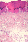
Eccrine angiomatous hamartomas are benign vascular and eccrine malformations often accompanied by hyperhidrosis or pain, increased eccrine glands, and aggregates of vessels. Verrucous hemangiomas are congenital vascular malformations presenting as unilateral grouped papules. Histologically, they show verrucous epidermal change and proliferation of capillaries in the dermis. We report a case of a 47-year-old woman with a red keratotic patch overlying a bluish plaque on the right sole, which had been present since birth. It was accompanied by pain and hyperhidrosis. Histologically, there were verrucous changes in the epidermis, numerous dilated capillaries in the papillary dermis, and increased eccrine glands with angiomatous foci in the deep dermis. The epithelial cells of the eccrine glands were positive for CEA, and the endothelial cells were positive for CD31 and GLUT-1. Eccrine angiomatous hamartomas have been reported in conjunction with other vascular tumors in only a few instances. We report an interesting case of an eccrine angiomatous hamartoma associated with a verrucous hemangioma.
Keywords: Eccrine angiomatous hamartoma; Verrucous hemangioma.
Figures


Similar articles
-
Eccrine angiomatous hamartoma, with verrocous hemangioma-like features: A case report.Int J Health Sci (Qassim). 2013 Jan;7(1):103-6. doi: 10.12816/0006027. Int J Health Sci (Qassim). 2013. PMID: 23559910 Free PMC article.
-
Eccrine angiomatous hamartoma with features resembling verrucous hemangioma.J Cutan Pathol. 2007 Dec;34 Suppl 1:68-70. doi: 10.1111/j.1600-0560.2007.00739.x. J Cutan Pathol. 2007. PMID: 17997743
-
Bilateral eccrine angiomatous hamartomas of the proximal interphalangeal joints.Dermatol Online J. 2023 Apr 15;29(2). doi: 10.5070/D329260767. Dermatol Online J. 2023. PMID: 37220283
-
[Congenital eccrine angiomatous hamartoma].Ann Dermatol Venereol. 1997;124(9):623-5. Ann Dermatol Venereol. 1997. PMID: 9739926 Review. French.
-
Eccrine angiomatous hamartoma: report of a case and literature review.J Am Acad Dermatol. 1999 Jul;41(1):109-11. doi: 10.1016/s0190-9622(99)70416-0. J Am Acad Dermatol. 1999. PMID: 10411421 Review.
Cited by
-
Eccrine Angiomatous Hamartoma in a Patient with Nevus Depigmentosus and Nevus Spilus.Indian J Dermatol. 2017 Jan-Feb;62(1):99-100. doi: 10.4103/0019-5154.198034. Indian J Dermatol. 2017. PMID: 28216737 Free PMC article. No abstract available.
-
Eccrine angiomatous hamartoma, with verrocous hemangioma-like features: A case report.Int J Health Sci (Qassim). 2013 Jan;7(1):103-6. doi: 10.12816/0006027. Int J Health Sci (Qassim). 2013. PMID: 23559910 Free PMC article.
-
Verrucous Hemangioma Treated with Electrocautery.Case Rep Dermatol. 2016 May 23;8(2):112-7. doi: 10.1159/000446100. eCollection 2016 May-Aug. Case Rep Dermatol. 2016. PMID: 27462218 Free PMC article.
-
Eccrine Angiomatous Hamartoma in an Adolescent.Case Rep Dermatol. 2015 Sep 5;7(3):233-6. doi: 10.1159/000439399. eCollection 2015 Sep-Dec. Case Rep Dermatol. 2015. PMID: 26500534 Free PMC article.
References
-
- Pelle MT, Pride HB, Tyler WB. Eccrine angiomatous hamartoma. J Am Acad Dermatol. 2002;47:429–435. - PubMed
-
- Lee HW, Han SS, Kang J, Lee MW, Choi JH, Moon KC, et al. Multiple mucinous and lipomatous variant of eccrine angiomatous hamartoma associated with spindle cell hemangioma: a novel collision tumor? J Cutan Pathol. 2006;33:323–326. - PubMed
-
- Galan A, McNiff JM. Eccrine angiomatous hamartoma with features resembling verrucous hemangioma. J Cutan Pathol. 2007;34(Suppl 1):68–70. - PubMed
-
- Tsuji T, Sawada H. Eccrine angiomatous hamartoma with verrucous features. Br J Dermatol. 1999;141:167–169. - PubMed
Publication types
LinkOut - more resources
Full Text Sources
Miscellaneous

