

Ecthyma Gangrenosum: A Rare Cutaneous Manifestation Caused by Stenotrophomonas maltophilia in a Leukemic Patient
- PMID: 20523829
- PMCID: PMC2861264
- DOI: 10.5021/ad.2009.21.4.389
Ecthyma Gangrenosum: A Rare Cutaneous Manifestation Caused by Stenotrophomonas maltophilia in a Leukemic Patient
Abstract
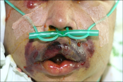
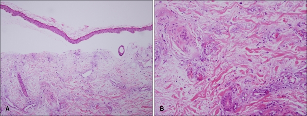
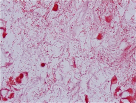
Ecthyma gangrenosum (EG) is a well-recognized cutaneous infection that most commonly affects immunocompromised patients. It typically occurs on the extremities, or in gluteal and perineal regions. Although Pseudomonas aeruginosa is the most well-known pathogen causing EG, other organisms have been reported to cause EG. Herein we report a rare case of ecthyma gangrenosum presenting as aggressive necrotic skin lesions in perioral and infraorbital areas in a 47-year-old patient with acute myelocytic leukemia after allogeneic bone marrow transplantation. It was caused by Stenotrophomonas maltophilia, which is an aerobic, gram-negative pathogen that has been associated only rarely with cutaneous disease. Blood culture and tissue culture were positive for S. maltophilia. Histological examination revealed numerous tiny bacilli in the dermis and perivascular area. Early recognition of skin lesions caused by S. maltophilia is important to decrease associated mortality in immunosuppressed patients.
Keywords: Ecthyma gangrenosum; Stenotrophomonas maltophilia.
Figures
Similar articles
-
Ecthyma gangrenosum: a rare manifestation of Stenotrophomonas maltophilia infection in acute myelogenous leukemia patient.IDCases. 2021 Oct 7;26:e01304. doi: 10.1016/j.idcr.2021.e01304. eCollection 2021. IDCases. 2021. PMID: 34703764 Free PMC article.
-
Ecthyma gangrenosum without bacteraemia in a leukaemic patient.Clin Exp Dermatol. 2001 Jul;26(5):395-7. doi: 10.1046/j.1365-2230.2001.00843.x. Clin Exp Dermatol. 2001. PMID: 11488824
-
Ecthyma gangrenosum: a rare cutaneous manifestation of a potentially fatal disease.Ann Otol Rhinol Laryngol. 2004 Jun;113(6):462-4. doi: 10.1177/000348940411300609. Ann Otol Rhinol Laryngol. 2004. PMID: 15224830
-
Nonpseudomonal ecthyma gangrenosum.J Am Acad Dermatol. 2004 May;50(5 Suppl):S114-7. doi: 10.1016/j.jaad.2003.09.019. J Am Acad Dermatol. 2004. PMID: 15097944 Review.
-
Ecthyma gangrenosum without bacteremia in a 54-year-old woman with heart transplant.Transpl Infect Dis. 2020 Aug;22(4):e13319. doi: 10.1111/tid.13319. Epub 2020 May 26. Transpl Infect Dis. 2020. PMID: 32396652 Review.
Cited by
-
Non healing leg ulcer infected with Stenotrophomonas maltophilia: first reported case from India.Int Wound J. 2013 Jun;10(3):356-8. doi: 10.1111/j.1742-481X.2012.00938.x. Epub 2012 Jan 31. Int Wound J. 2013. PMID: 22289105 Free PMC article.
-
Fatal Oculocutaneous Ecthyma Gangrenosum in Human Immunodeficiency Virus/Acquired Immunodeficiency Syndrome: Case Report and Review of the Literature.J Glob Infect Dis. 2019 Jan-Mar;11(1):43-46. doi: 10.4103/jgid.jgid_54_18. J Glob Infect Dis. 2019. PMID: 30814835 Free PMC article.
-
Empiric intralesional tumescent drug delivery of antimicrobials effectively treated a painful necrotizing skin infection.JAAD Case Rep. 2024 Mar 26;50:40-43. doi: 10.1016/j.jdcr.2024.03.008. eCollection 2024 Aug. JAAD Case Rep. 2024. PMID: 39036615 Free PMC article. No abstract available.
-
A Challenging Cutaneous Lesion in a Patient With Chronic Idiopathic Neutropenia.Cureus. 2022 Jan 14;14(1):e21225. doi: 10.7759/cureus.21225. eCollection 2022 Jan. Cureus. 2022. PMID: 35186525 Free PMC article.
-
Ecthyma gangrenosum: a rare manifestation of Stenotrophomonas maltophilia infection in acute myelogenous leukemia patient.IDCases. 2021 Oct 7;26:e01304. doi: 10.1016/j.idcr.2021.e01304. eCollection 2021. IDCases. 2021. PMID: 34703764 Free PMC article.
References
-
- James WD, Berger TG, Elston DM. Andrews' diseases of the skin: clinical dermatology. 10th ed. Philadelphia: Saunders Elsevier; 2006. pp. 271–272.
-
- McKee PH, Calonje E, Granter SR. Pathology of the skin: with clinical correlations. 3rd ed. Philadelphia: Elsevier Mosby; 2005. pp. 869–871.
-
- Downey DM, O'Bryan MC, Burdette SD, Michael JR, Saxe JM. Ecthyma gangrenosum in a patient with toxic epidermal necrolysis. J Burn Care Res. 2007;28:198–202. - PubMed
-
- Pandit AM, Siddaramappa B, Choudhary SV, Manjunathswamy BS. Ecthyma gangrenosum in a new born child. Indian J Dermatol Venereol Leprol. 2003;69:52–53. - PubMed
-
- Reich HL, Williams Fadeyi D, Naik NS, Honig PJ, Yan AC. Nonpseudomonal ecthyma gangrenosum. J Am Acad Dermatol. 2004;50(5 Suppl):S114–S117. - PubMed
Publication types
LinkOut - more resources
Full Text Sources
