. 2010 May 27;6(5):e1000973.
doi: 10.1371/journal.pgen.1000973.

Sgs1 and exo1 redundantly inhibit break-induced replication and de novo telomere addition at broken chromosome ends

Affiliations

Sgs1 and exo1 redundantly inhibit break-induced replication and de novo telomere addition at broken chromosome ends

John R Lydeard et al. PLoS Genet. .

Abstract

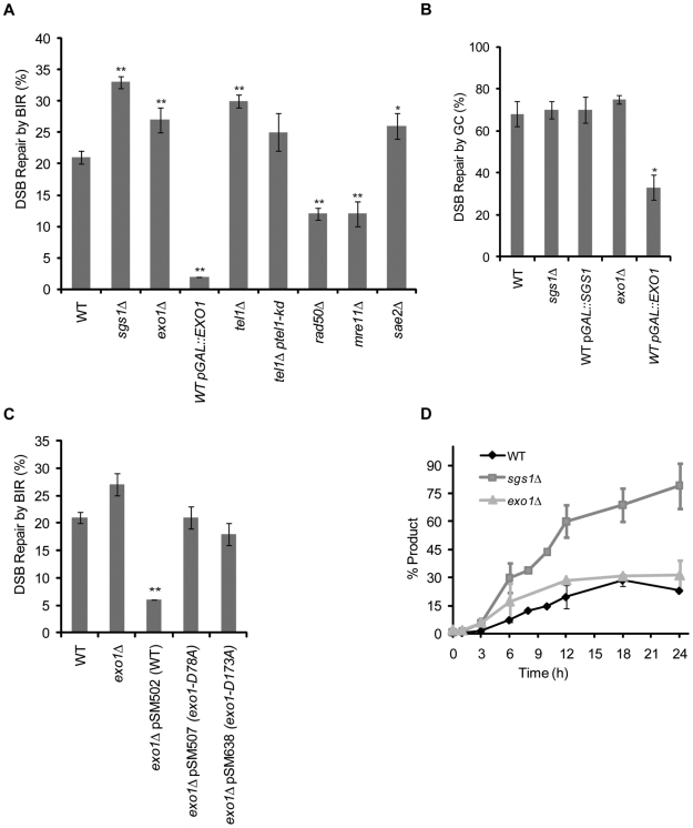
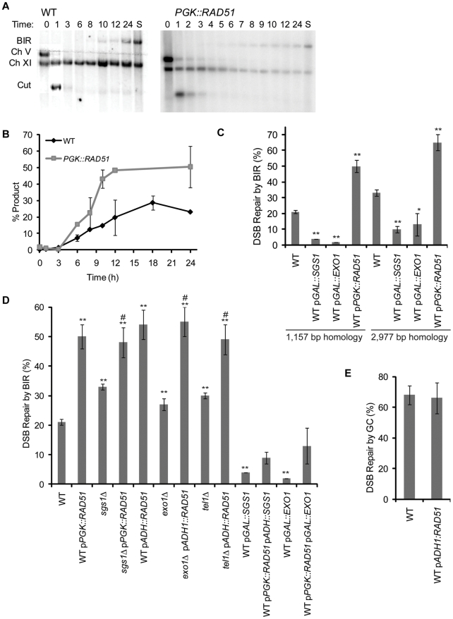
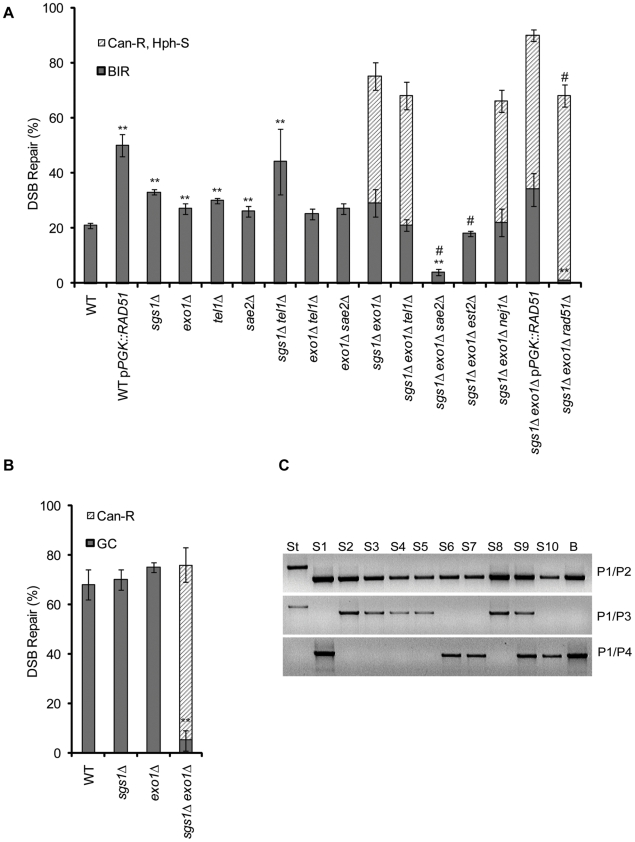
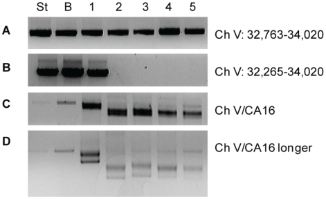
In budding yeast, an HO endonuclease-inducible double-strand break (DSB) is efficiently repaired by several homologous recombination (HR) pathways. In contrast to gene conversion (GC), where both ends of the DSB can recombine with the same template, break-induced replication (BIR) occurs when only the centromere-proximal end of the DSB can locate homologous sequences. Whereas GC results in a small patch of new DNA synthesis, BIR leads to a nonreciprocal translocation. The requirements for completing BIR are significantly different from those of GC, but both processes require 5' to 3' resection of DSB ends to create single-stranded DNA that leads to formation of a Rad51 filament required to initiate HR. Resection proceeds by two pathways dependent on Exo1 or the BLM homolog, Sgs1. We report that Exo1 and Sgs1 each inhibit BIR but have little effect on GC, while overexpression of either protein severely inhibits BIR. In contrast, overexpression of Rad51 markedly increases the efficiency of BIR, again with little effect on GC. In sgs1Delta exo1Delta strains, where there is little 5' to 3' resection, the level of BIR is not different from either single mutant; surprisingly, there is a two-fold increase in cell viability after HO induction whereby 40% of all cells survive by formation of a new telomere within a few kb of the site of DNA cleavage. De novo telomere addition is rare in wild-type, sgs1Delta, or exo1Delta cells. In sgs1Delta exo1Delta, repair by GC is severely inhibited, but cell viability remains high because of new telomere formation. These data suggest that the extensive 5' to 3' resection that occurs before the initiation of new DNA synthesis in BIR may prevent efficient maintenance of a Rad51 filament near the DSB end. The severe constraint on 5' to 3' resection, which also abrogates activation of the Mec1-dependent DNA damage checkpoint, permits an unprecedented level of new telomere addition.


Conflict of interest statement

The authors have declared that no competing interests exist.

Figures

Figure 1
Figure 1. Experimental systems of break-induced replication (BIR) and gene conversion (GC).
(A) In the experimental system to study BIR, an HPHMX marked HO cut site (gray bar) is integrated into the CAN1 gene on Ch V, deleting the 3′ end portion of the gene, the remaining sequences are represented as CA. The AN1 donor sharing 1,157 bp homology with CAN1 is integrated into Ch XI. PCR with primers P1 and P2 monitors the initiation of new DNA synthesis while PCR with primers P1 and P4 detects synthesis past the AN1 sequences, specifc to the donor sequences on Ch XI. Southern blot analysis of AvaI-digested (marked by “A”) DNA probed with CAN1 sequences monitors extension of the BIR fork. Completion of BIR is monitored by Pulse-field gel electrophoresis (PFGE) followed by Southern blot analysis using the MCH2 sequences that are duplicated when the entire donor chromosome arm is copied. (B) In the experimental system to study ectopic GC. A galactose inducible HO endonuclease generates a DSB within the CAN1 locus (disrupted by URA3 creating a 376 bp gap) on Ch V. An additional 2,404 bp of homologous sequences to the gene conversion donor sequences found on Ch XI are distal to the cut site and are denoted as “1.” PCR with primers P1 and P2 monitors both the starting strain and repair into the CAN1 sequences. PCR with primers P1 and P3 monitors repair by GC in which the distal end of the break is retained.
Figure 2
Figure 2. Sgs1 and Exo1 negatively regulate BIR.
(A) Efficiency of BIR in cells as measured by viability following a DSB. (B) Efficiency of GC in cells as measured by viability following a DSB. (C) Efficiency of BIR in wild type (WT), exo1Δ, overexpression of EXO1 and overexpression of EXO1 nuclease-dead alleles measured by viability following a DSB. For (A–C), data are the mean ±standard error of the mean. Values marked with asterics are statistically significant (*represents p<0.05, ** represents p<0.01 compared to wild type). (D) The kinetics of repair are shown for PCR of BIR induced in cycling WT, sgs1Δ and exo1Δ cells amplified with P1 and P2 primer set labeled as “CAN1” and the standard FLO9 locus of. Data are the mean ±standard deviation.
Figure 3
Figure 3. Overexpression of RAD51 increases the kinetics and efficiency of BIR.
(A) Southern blot analysis of the kinetics of repair product in wild type and pPGK::RAD51 cycling cells as indicated in Figure 1A. Lane S contains DNA from a colony where BIR occurred. (B) Kinetics of repair are shown for PCR of BIR induced in cycling wild type (WT) and pPGK::RAD51 cells. Data are the mean ±data range. (C) Efficiency of BIR in cells as measured by viability following a DSB in a BIR assay with increased homology (2,977 bp homology). Data from Figure 2A and Figure 3D (1,157 bp homology strain) are shown for comparison. Data are the mean ±s.e.m. Values marked with asterics are statistically significant (*represents p < 0.05, ** represents p < 0.01 compared to wild type). (D) Efficiency of BIR in strains graphed in Figure 2 also carrying either pPGK::RAD51 or pADH::RAD51 as measured by viability following a DSB. Data are the mean ±s.e.m. Values marked with asterics or number sign are statistically significant (*represents p < 0.05, ** represents p < 0.01 compared to wild type. # represents p < 0.05 to the corresponding single mutant). (E) Efficiency of GC in WT and pPGK::RAD51 as measured by viability following a DSB.
Figure 4
Figure 4. The effect of sgs1Δ exo1Δ on the viability and repair product in BIR and GC.
(A) The viability and phenotypic characterization of wild type (WT), sgs1Δ, exo1Δ, tel1Δ, sgs1Δsgs1Δ exo1Δ, pPGK::RAD51 and indicated double and triple mutant combination cells following a DSB in the BIR assay. BIR colonies (CanS HphS) represent those that have repaired the DSB by BIR while CanR HphS colonies represent those that have a truncated chromosome. Data are the mean ±s.e.m. Values marked with asterics or number sign are statistically significant (*represents p < 0.05, ** represents p < 0.01 compared to wild type BIR. # represents p < 0.05 to the sgs1Δ exo1Δ CanR HphS colonies). (B) The viability and phenotypic characterization of cells following a DSB in the GC assay. HR colonies (CanS) represent those that have repaired by Homologus Recombination (either BIR or GC) while CanR colonies represent those that have a truncated chromosome. Data are the mean ±s.e.m. ** represents p < 0.01 compared to wild type. (C) Repair of CanS colonies in the GC assay as monitored by PCR. Included are the starting GC strain (ST), ten CanS colonies (S1–S10) and a colony that has repaired by BIR (B). PCR with primers P1 and P2 detects the starting band and shift to smaller size upon repair into the CAN1 sequences if repair occurs either by GC or BIR. PCR of primers P1 and P3 monitors retention of the distal end of the DSB and is indicative of repair by GC. PCR with primers P1 and P4 monitors repair specific to BIR (see Figure 1).
Figure 5
Figure 5. Characterization of CanR HPHS sgs1Δ exo1Δ colonies in the BIR strain by PFGE.
(A) Ethidium bromide-stained agarose gel PFGE gel of sgs1Δ exo1Δ colonies that have repaired the DSB. Included are the ladder (L), starting strain prior to DSB induction (ST), CanS HPHS colony that has repaired by BIR (B), and twelve CanR HPHS colonies (1–12). Arrows indicate additional uncharacterized chromosomal fragments. (B) Southern blot analysis of (5A) by hybridization with a probe for MCH2 that normally lies 6 kb from the telomere on Ch XI (See Figure 1). (C) The blot was stripped and Southern blot analysis was performed by hybridization with a probe for CAN1 that normally lies 33 kb from the telomere on Ch V and is 1 kb proximal to the HO cut site (See Figure 1). (D) The blot was stripped and Southern blot analysis was performed by hybridization with a probe for NPR2 that normally lies 36 kb from the telomere on Ch V and is 4 kb proximal to the HO cut site (See Figure 1). (E) Southern blot analysis was performed on (5D) by hybridization with a probe for PRB1 that normally lies 40 kb from the telomere on Ch V and is 8 kb proximal to the HO cut site (See Figure 1).
Figure 6
Figure 6. Marking of the breakpoint and detection of de novo telomere formation by PCR in sgs1Δ exo1Δ CanR HphS cells.
From the BIR assay. (A) PCR analysis of a starting strain prior to DSB induction (ST), CanS HphS colony that has repaired by BIR (B), and five CanR HPHS colonies (1–5) with primers that amplify sequences (Ch V 32,763–34,020) approximately 750 bp proximal to the break. (B) PCR with primers that amplify sequences (Ch V 32,265–34,020) approximately 250 bp proximal to the break. (C) PCR with a Ch V-specific primer that amplifies all colonies indicated and primer CA16, a telomere-specific primer. (D) PCR product from 6C ran longer an agarose gel to better display the laddered PCR product indicative of de novo telomere formation in samples 1–5.

Similar articles

Cited by

References

    1. San Filippo J, Sung P, Klein H. Mechanism of eukaryotic homologous recombination. Annu Rev Biochem. 2008;77:229–257. - PubMed
    1. Agmon N, Pur S, Liefshitz B, Kupiec M. Analysis of repair mechanism choice during homologous recombination. Nucleic Acids Res. 2009;37:5081–5092. - PMC - PubMed
    1. Krogh BO, Symington LS. Recombination proteins in yeast. Annu Rev Genet. 2004;38:233–271. - PubMed
    1. Pâques F, Haber JE. Multiple pathways of recombination induced by double-strand breaks in Saccharomyces cerevisiae. Microbiol Mol Biol Rev. 1999;63:349–404. - PMC - PubMed
    1. Llorente B, Smith CE, Symington LS. Break-induced replication: what is it and what is it for? Cell Cycle. 2008;7:859–864. - PubMed

Publication types