. 2010 May 27;6(5):e1000921.
doi: 10.1371/journal.ppat.1000921.

Effective, broad spectrum control of virulent bacterial infections using cationic DNA liposome complexes combined with bacterial antigens

Affiliations

Effective, broad spectrum control of virulent bacterial infections using cationic DNA liposome complexes combined with bacterial antigens

Robin Ireland et al. PLoS Pathog. .

Abstract

Protection against virulent pathogens that cause acute, fatal disease is often hampered by development of microbial resistance to traditional chemotherapeutics. Further, most successful pathogens possess an array of immune evasion strategies to avoid detection and elimination by the host. Development of novel, immunomodulatory prophylaxes that target the host immune system, rather than the invading microbe, could serve as effective alternatives to traditional chemotherapies. Here we describe the development and mechanism of a novel pan-anti-bacterial prophylaxis. Using cationic liposome non-coding DNA complexes (CLDC) mixed with crude F. tularensis membrane protein fractions (MPF), we demonstrate control of virulent F. tularensis infection in vitro and in vivo. CLDC+MPF inhibited bacterial replication in primary human and murine macrophages in vitro. Control of infection in macrophages was mediated by both reactive nitrogen species (RNS) and reactive oxygen species (ROS) in mouse cells, and ROS in human cells. Importantly, mice treated with CLDC+MPF 3 days prior to challenge survived lethal intranasal infection with virulent F. tularensis. Similarly to in vitro observations, in vivo protection was dependent on the presence of RNS and ROS. Lastly, CLDC+MPF was also effective at controlling infections with Yersinia pestis, Burkholderia pseudomallei and Brucella abortus. Thus, CLDC+MPF represents a novel prophylaxis to protect against multiple, highly virulent pathogens.


Conflict of interest statement

A provisional patent for CLDC+MPF has been filed by NIH on behalf of Catharine M. Bosio, John T. Belise and Jeff Fairman.

Figures
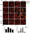
Figure 1
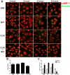
Figure 1. CLDC+MPF mediated control of F. tularensis replication in mouse macrophages.
Mouse macrophages were treated with D5W (untreated), MPF, CLDC, or CLDC+MPF 18 h prior to infection. Four, 8, 12 and 24 h after infection phagocytosis and intracellular replication of F. tularensis was monitored by microscopy. Exposure to CLDC, MPF or CLDC+MPF did not impact the number of cells infected (A and C) nor the number of bacteria entering each cell (B). MPF and CLDC treatment alone significantly increased the number SchuS4 infected cells 24 h after infection compared to untreated controls (* = p<0.01) (A and C). In contrast, CLDC+MPF treated cultures had fewer infected cells within 8 h of infection through 24 h after infection (** = p<0.01) (A and C). White arrows indicate intracellular SchuS4. Data is representative of four experiments. Error bars represent SEM.
Figure 2
Figure 2. CLDC+MPF mediated control of F. tularensis replication in human macrophages.
Human macrophages were treated with D5W (untreated), MPF, CLDC, or CLDC+MPF 18 h prior to infection. Four, 8, 12 and 24 h after infection phagocytosis and intracellular replication of F. tularensis was monitored by microscopy. Exposure to CLDC, MPF or CLDC+MPF did not impact the number of cells infected (A and C) nor the number of bacteria entering each cell (B). CLDC+MPF treated cultures had fewer infected cells within 12 h of infection and through 24 h after infection (* = p<0.01) (A and C). White arrows indicate intracellular SchuS4. Data is representative of four experiments. Error bars represent SEM.
Figure 3
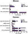
Figure 3. Induction of RNS and ROS genes by CLDC+MPF.
Mouse and human macrophages were treated with D5W, CLDC, MPF or CLDC+MPF. After 16 and 12 h, respectively, gene expression was monitored by quantitative RT-PCR. Change in the expression of the indicated genes is represented as fold change over D5W treated controls. Data is representative of three experiments.
Figure 4
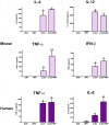
Figure 4. CLDC+MPF induced production of pro-inflammatory cytokines in macrophages.
Mouse and human macrophages were treated with D5W (untreated), MPF, CLDC, or CLDC+MPF and 24 h later supernatants were assessed for cytokines by ELISA. In mouse cells, CLDC and CLDC+MPF induced significantly more IL-6, IL-12 and IFN-β compared to D5W or MPF treated cells (* = p<0.01). Whereas, CLDC+MPF induced significantly more TNF-α compared to all other groups (** = p<0.01). In human cells CLDC and CLDC+MPF induced significantly more TNF-α compared to D5W or MPF treated cells (* = p<0.01). Whereas CLDC+MPF induced significantly more IL-6 compared to all other groups (** = p<0.01). Data is representative of four experiments. Error bars represent SEM.
Figure 5
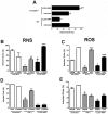
Figure 5. CLDC+MPF control of F. tularensis is dependent on RNS and ROS.
(A) Macrophages from wild type or nos2/gp91−/− mice were treated with D5W (untreated) or CLDC+MPF for 18 h followed by infection with SchuS4. Intracellular replication was monitored by microscopy and the percent infected cells is depicted for each group. CLDC+MPF significantly reduced the number of SchuS4 infected wild type cells (* = p<0.01), whereas CLDC+MPF treatment of nos2/gp91−/− significantly increased the number of infected macrophages (** = p<0.01). (B and C) Wild type mouse macrophages were treated with D5W (untreated), IFN-γ or CLDC+MPF in the presence or absence of L-NMMA (B) or NAC (C) and then infected with SchuS4. IFN-γ and CLDC+MPF significantly lowered SchuS4 infected cells (* = p<0.05) compared to untreated controls. Addition of L-NMMA and NAC significantly increased the number of infected macrophages compared to IFN-γ (** = p<0.05) or CLDC+MPF (*** = p<0.01) treated controls. (D and E) Human macrophages were treated with D5W (untreated), IFN-γ or CLDC+MPF in the presence or absence of L-NMMA (D) or NAC (E) and then infected with SchuS4. IFN-γ and CLDC+MPF significantly lowered SchuS4 infected cells (* = p<0.05) compared to untreated controls. Addition of L-NMMA significantly increased the number of infected macrophages compared to IFN-γ (** = p<0.05), but not CLDC+MPF treated controls. Addition of NAC significantly increased the number of infected macrophages compared to CLDC+MPF (** = p<0.01), but not IFN-γ, treated controls.
Figure 6
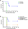
Figure 6. CLDC+MPF enhanced survival of SchuS4 infection in vivo.
(A) Mice (n = 10 per group) were treated with CLDC+MPF i.v. 3 days prior to intranasal challenge with SchuS4. Significantly more CLDC+MPF treated mice survived infection compared to untreated controls (p = 0.0027). (B) Wild type (WT) or nos2/gp91−/−mice (n = 5 per group) were treated with CLDC+MPF 3 days prior to challenge with SchuS4. Sixty percent of CDLC+MPF treated WT mice survived infection, whereas significantly fewer nos2/gp91−/− survived (p = 0.0385). Data are representative of two experiments.
Figure 7
Figure 7. CLDC+MPF mediated protection against unrelated bacterial pathogens.
Human macrophages were untreated (D5W) or treated with CLDC+MPF 18 h prior to infection followed by infection with B. pseudomallei, Y. pestis or B. abortus, At the indicated time points cells were analyzed for intracellular bacteria by microscopy. CDLC+MPF significantly reduced the number of infected cells and the number of bacteria per cell regardless of the species of infecting bacteria (* = p<0.05). Data is representative of three experiments. Error bars represent SEM.

Similar articles

Cited by

References

    1. Conlan JW, Chen W, Shen H, Webb A, KuoLee R. Experimental tularemia in mice challenged by aerosol or intradermally with virulent strains of Francisella tularensis: bacteriologic and histopathologic studies. Microb Pathog. 2003;34:239–248. - PubMed
    1. Saslaw S, Eigelsbach HT, Prior JA, Wilson HE, Carhart S. Tularemia vaccine study. II. Respiratory challenge. Arch Intern Med. 1961;107:702–714. - PubMed
    1. Sawyer WD, Dangerfield HG, Hogge AL, Crozier D. Antibiotic prophylaxis and therapy of airborne tularemia. Bacteriol Rev. 1966;30:542–550. - PMC - PubMed
    1. Cross JT, Jacobs RF. Tularemia: treatment failures with outpatient use of ceftriaxone. Clin Infect Dis. 1993;17:976–980. - PubMed
    1. Overholt EL, Tigertt WD, Kadull PJ, Ward MK, Charkes ND, et al. An analysis of forty-two cases of laboratory-acquired tularemia. Treatment with broad spectrum antibiotics. Am J Med. 1961;30:785–806. - PubMed

Publication types

MeSH terms