. 2010 May 27;6(5):e1000971.
doi: 10.1371/journal.pgen.1000971.

Aging and chronic sun exposure cause distinct epigenetic changes in human skin

Affiliations

Aging and chronic sun exposure cause distinct epigenetic changes in human skin

Elke Grönniger et al. PLoS Genet. .

Abstract

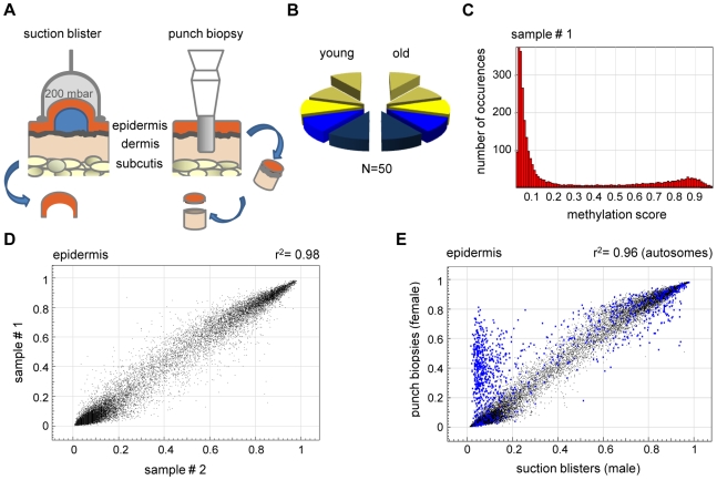
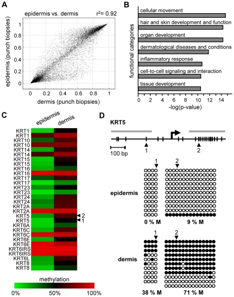
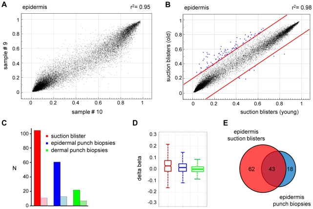
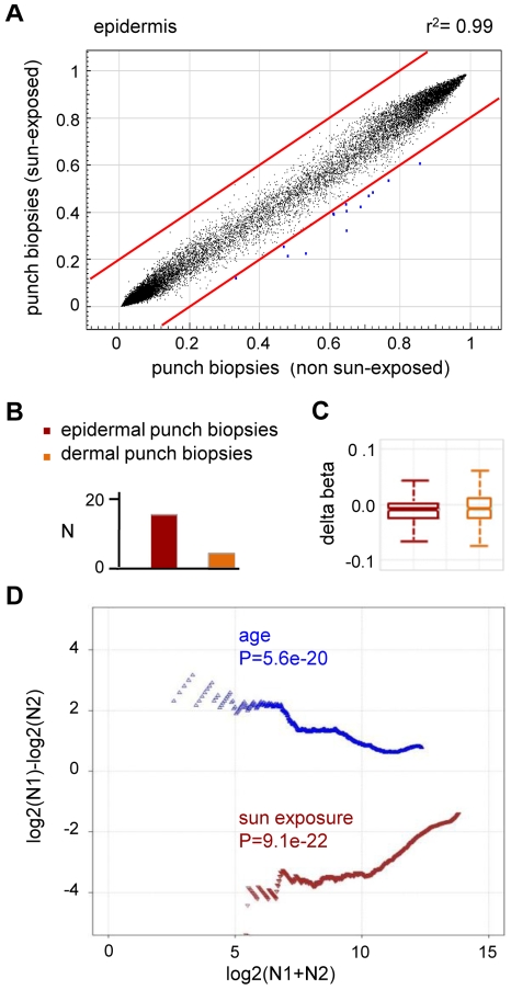
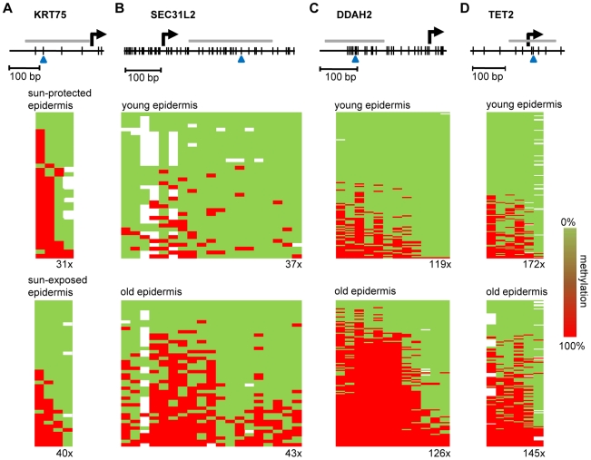
Epigenetic changes are widely considered to play an important role in aging, but experimental evidence to support this hypothesis has been scarce. We have used array-based analysis to determine genome-scale DNA methylation patterns from human skin samples and to investigate the effects of aging, chronic sun exposure, and tissue variation. Our results reveal a high degree of tissue specificity in the methylation patterns and also showed very little interindividual variation within tissues. Data stratification by age revealed that DNA from older individuals was characterized by a specific hypermethylation pattern affecting less than 1% of the markers analyzed. Interestingly, stratification by sun exposure produced a fundamentally different pattern with a significant trend towards hypomethylation. Our results thus identify defined age-related DNA methylation changes and suggest that these alterations might contribute to the phenotypic changes associated with skin aging.


Conflict of interest statement

The authors have declared that no competing interests exist.

Figures

Figure 1
Figure 1. High interindividual similarity between DNA methylation patterns from human epidermis.
(A) Procedures for skin sample preparation. Suction blisters were induced on the forearms of healthy volunteers and suction blister roofs were taken as sources for epidermal DNA (left panel). Punch biopsies were obtained from the outer forearm (sun-exposed) and inner arm (sun-protected) and separated into epidermal and dermal layers by dispase II treatment. (B) Schematic outline of the sample groups analyzed in this study. Epidermis (yellow) and dermis (blue) samples were obtained from sun-exposed (bright colors) and sun-protected (shaded colors) skin areas, either as suction blisters (elevated) or as punch biopsies. Each segment represents 5 samples. (C) Representative methylation profile of a human epidermis sample. Most markers were found to be unmethylated (beta<0.2), and a smaller group of markers was found to be highly methylated (beta>0.8). (D) DNA methylation profiles were compared between suction blister samples from two independent young donors. The results show a very high similarity with a correlation coefficient of r2 = 0.98. (E) Epidermal DNA methylation profiles were compared between two independent studies. Suction blister samples were obtained from male volunteers, punch biopsy samples were obtained from female individuals. Markers with major variations were almost completely localized to the X-chromosome (marked in blue), and can thus be attributed to the known hypermethylation of X-chromosomal loci in females.
Figure 2
Figure 2. Distinct DNA methylation patterns of human epidermis and dermis.
(A) Comparison of average DNA methylation profiles between 20 epidermal and 20 dermal punch biopsy samples. A substantial number of genes show major methylation differences. (B) Ingenuity Pathway Analysis (www.ingenuity.com) of markers with differential (Δ(beta)≥0.2) methylation values in epidermis and dermis. The plot shows the seven most significantly overrepresented functional categories, which are closely associated with (skin) tissue development. Differential methylation of genes associated with “cellular movement” conceivably reflects the prominent differences in the tissue organization between epidermis and dermis. (C) Heatmap of Keratin gene markers with increased (Δ(beta)≥0.2) dermal methylation levels of at least one marker per gene. (D) Validation of differential KRT5 methylation by bisulfite sequencing. Top: Structure of the KRT5 promoter region. CpG dinucleotides are shown as vertical lines, PCR amplicons are shown as grey horizontal bars, the transcription start site (TSS) is indicated by an arrow, numbered arrowheads highlight the CpG sites represented on the Infinium array. Bottom: Bisulfite sequencing results. Each row represents one sequence read, black circles indicate methylated CpG dinucleotides, white circles indicate unmethylated CpG dinucleotides.
Figure 3
Figure 3. Age-related hypermethylation in human epidermis samples.
(A) DNA methylation profiles were compared between epidermal blister samples from two independent old donors. The results show a high similarity with a correlation coefficient of r2 = 0.95. (B) Average epidermal DNA methylation profiles of the study population were compared between old and young epidermis samples. The results reveal a distinct trend towards age-associated hypermethylation. (C) Barplot illustrating the results from an independent statistical analysis of the array data. The number of substantially hypermethylated (Δ(beta)≥0.2, P(BH)<0.01) markers is shown in dark colors, the number of substantially hypomethylated (Δ(beta)≤−0.2, P(BH)<0.01) markers is shown in light colors. (D) Boxplot illustrating age-related methylation changes in human epidermis and dermis samples. (E) A comparison of substantially hypermethylated markers from two independent sets of epidermis samples (suction blisters and punch biopsies) reveals a large overlap of 43 commonly hypermethylated markers.
Figure 4
Figure 4. Sun exposure-related hypomethylation.
(A) Average epidermal DNA methylation profiles of the study population were compared between 10 sun-exposed and 10 non-exposed epidermis samples. The results reveal a distinct trend towards sun exposure-related hypomethylation. (B) Barplot illustrating the results from an independent statistical analysis of the array data. Bars indicate the number of substantially hypomethylated (Δ(beta)≤−0.2, P(BH)<0.01) markers. (C) Boxplot illustrating sun exposure-related methylation changes in human epidermis and dermis samples. (D) RS (ratio-over-sum) plot illustrating global age-related (blue) and sun exposure-related (brown) methylation shifts in epidermis samples. Points were plotted after Benjamini-Hochberg correction and P-values were obtained after comparisons to Gaussian distributions of randomly generated methylation differences.
Figure 5
Figure 5. Validation of methylation changes by deep bisulfite sequencing.
Schematic outline of the promoter regions of (A) KRT75 (B) SEC31L2, (C) DDAH2, and (D) TET2. Vertical lines represent individual CpG dinucleotides, blue arrowheads indicate CpG markers represented on the array. PCR amplification was performed on equimolar sample pools. PCR amplicons for sequencing are shown as grey horizontal bars, sequencing results are shown as heatmaps. Each row represents one sequence read, individual red boxes represent methylated CpG dinucleotides, green boxes represent unmethylated CpG dinucleotides, sequencing gaps are shown in white. Sequencing coverage ranged from 31x to 172x, as indicated. The deamination efficiency was >99% for all samples analyzed.

Similar articles

Cited by

References

    1. Fraga MF, Esteller M. Epigenetics and aging: the targets and the marks. Trends Genet. 2007;23:413–418. - PubMed
    1. Bird A. Perceptions of epigenetics. Nature. 2007;447:396–398. - PubMed
    1. Suzuki MM, Bird A. DNA methylation landscapes: provocative insights from epigenomics. Nat Rev Genet. 2008;9:465–476. - PubMed
    1. Kouzarides T. Chromatin modifications and their function. Cell. 2007;128:693–705. - PubMed
    1. Esteller M. CpG island hypermethylation and tumor suppressor genes: a booming present, a brighter future. Oncogene. 2002;21:5427–5440. - PubMed

Publication types