. 2010 Oct;53(10):2167-76.
doi: 10.1007/s00125-010-1809-6. Epub 2010 Jun 5.

Adaptive changes in pancreatic beta cell fractional area and beta cell turnover in human pregnancy

Affiliations

Adaptive changes in pancreatic beta cell fractional area and beta cell turnover in human pregnancy

A E Butler et al. Diabetologia. 2010 Oct.

Abstract

Aims/hypothesis: We sought to establish the extent and basis for adaptive changes in beta cell numbers in human pregnancy.

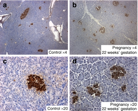
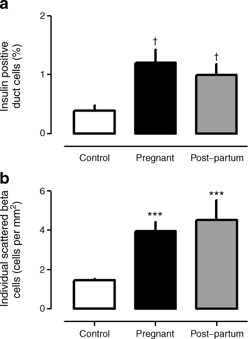
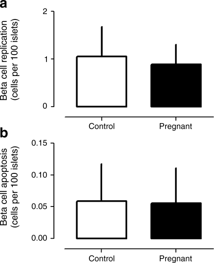
Methods: Pancreas was obtained at autopsy from women who had died while pregnant (n = 18), post-partum (n = 6) or were not pregnant at or shortly before death (controls; n = 20). Pancreases were evaluated for fractional pancreatic beta cell area, islet size and islet fraction of beta cells, beta cell replication (Ki67) and apoptosis (TUNEL), and indirect markers of beta cell neogenesis (insulin-positive cells in ducts and scattered beta cells in pancreas).

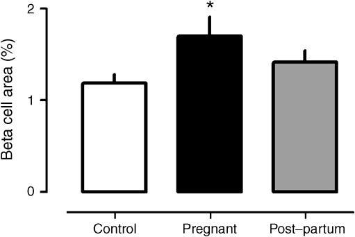
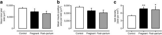
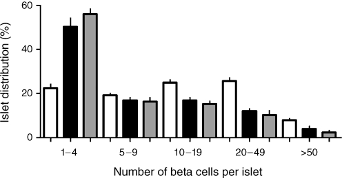
Results: The pancreatic fractional beta cell area was increased by approximately 1.4-fold in human pregnancy, with no change in mean beta cell size. In pregnancy there were more small islets rather than an increase in islet size or beta cells per islet. No increase in beta cell replication or change in beta cell apoptosis was detected, but duct cells positive for insulin and scattered beta cells were increased with pregnancy.

Conclusions/interpretation: The adaptive increase in beta cell numbers in human pregnancy is not as great as in most reports in rodents. This increase in humans is achieved by increased numbers of beta cells in apparently new small islets, rather than duplication of beta cells in existing islets, which is characteristic of pregnancy in rodents.


Figures

Fig. 1
Fig. 1
The mean fractional pancreatic beta cell area in non-pregnant controls, and in women who were pregnant or in the post-partum state. During pregnancy the mean fractional beta cell area was 1.4-fold increased (*p < 0.05) compared with the controls
Fig. 2
Fig. 2
Mean islet size (a), mean area per islet positive for insulin (b) and mean islet density (c) in non-pregnant controls, and in women who were pregnant or in the post-partum state; values determined by evaluating ten prominent islets per woman. During pregnancy and post-partum the mean islet size and the mean insulin area per islet were decreased (*p < 0.05) compared with non-pregnant controls. Islet density (c) was increased (**p < 0.01 pregnant; *p < 0.05 post-partum)
Fig. 3
Fig. 3
Sections of pancreas from control women (a, c) and pregnant women at 20 weeks (b) and 22 weeks (d) of gestation. Images were generated at low power (4×) (a, b) and at higher power (20×) (c, d), with samples stained for insulin (brown) and counterstained by haematoxylin. While a range of islet sizes is apparent on cross-section in both cases, the abundance of small islets is increased in the pregnant women
Fig. 4
Fig. 4
The frequency distribution of beta cells per islet on section in non-pregnant controls (white bars), and in women who were pregnant (black bars) or in the post-partum state (grey bars). There was a marked shift towards small islets in pregnancy and in the post-partum state compared with the non-pregnant state. This shift is the opposite to that observed in pregnant rodents, in which more abundant large islets have been detected
Fig. 5
Fig. 5
The mean percentage of pancreatic duct cells positive for insulin (a) and the abundance of single scattered beta cells (b) in pregnant controls, and in women who were pregnant or in the post-partum state. These indices, which have been used previously as indirect measures of beta cell neogenesis, were both increased with pregnancy, remaining so post-partum. ***p < 0.001 vs control; p < 0.005 vs control
Fig. 6
Fig. 6
The mean frequency of beta cell replication by Ki67 (a) and apoptosis by TUNEL (b) in non-pregnant controls and in pregnant women. As previously reported in humans, both indices are infrequent implying relatively slow beta cell turnover. No detectable change in either was observed in pregnancy. Given the low frequency of these measures and the limited pancreatic tissue available per woman, data were insufficient to allow reliable documentation of frequency of these variables in the six post-partum women

Comment in

Similar articles

Cited by

References

    1. Gepts W. Pathologic anatomy of the pancreas in juvenile diabetes mellitus. Diabetes. 1965;14:619–633. - PubMed
    1. Butler AE, Janson J, Bonner-Weir S, Ritzel R, Rizza RA, Butler PC. Beta-cell deficit and increased beta-cell apoptosis in humans with type 2 diabetes. Diabetes. 2003;52:102–110. doi: 10.2337/diabetes.52.1.102. - DOI - PubMed
    1. Parsons JA, Brelje TC, Sorenson RL. Adaptation of islets of Langerhans to pregnancy: increased islet cell proliferation and insulin secretion correlates with the onset of placental lactogen secretion. Endocrinology. 1992;130:1459–1466. doi: 10.1210/en.130.3.1459. - DOI - PubMed
    1. Scaglia L, Smith FE, Bonner-Weir S. Apoptosis contributes to the involution of beta cell mass in the post partum rat pancreas. Endocrinology. 1995;136:5461–5468. doi: 10.1210/en.136.12.5461. - DOI - PubMed
    1. Karnik SK, Chen H, McLean GW, et al. Menin controls growth of pancreatic beta-cells in pregnant mice and promotes gestational diabetes mellitus. Science. 2007;318:806–809. doi: 10.1126/science.1146812. - DOI - PubMed

Publication types