

doi: 10.1021/jm100086u.
Host-guest chemistry of the peptidoglycan
Affiliations
- PMID: 20524613
- PMCID: PMC2908483
- DOI: 10.1021/jm100086u
Item in Clipboard
Host-guest chemistry of the peptidoglycan
J Med Chem.
.
No abstract available
Figures
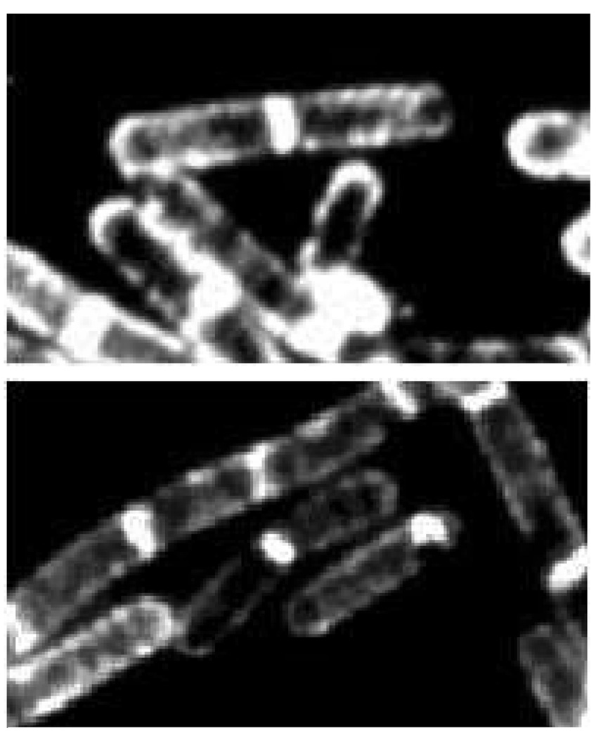
Visualization of the external peptidoglycan surface of Gram-positive bacteria by the use of fluorescently-labeled peptidoglycan-binding antibiotics. These fluorescent microscopy images, reproduced from the study by Tiyanont et al. Proc. Natl Acad. Sci. U. S. A. 2006, 103, 11033–11038 © 2006 National Academy of Sciences, show rod-shaped B. subtilis (the bacterium is approximately 2 µm in length) bacteria stained with fluorescein-labeled ramoplanin. The peptidoglycan is intensely stained at the newly forming division septum, and to a lesser extent on the sidewalls and the old pole. These locations coincide with the sub-cellular locations of Lipid II, the key biosynthetic precursor of the peptidoglycan (see Scheme 1). The sidewall staining is suggestive of a helical pattern for peptidoglycan growth during sidewall elongation.
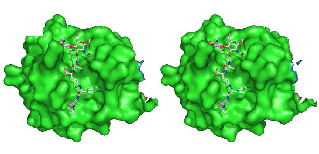
Computational stereo structure of the [NAG-NAM]2 structure (see Scheme 2) in complex with the human PGRP-1® peptidodglycan recognition protein, based on an X-ray structure of a smaller synthetic fragment bound to the protein (PDB Code 2EAX). The approximate diameter of the protein is 4 nm. The perspective shown for the protein orients the peptidoglycan cleft from top to bottom.
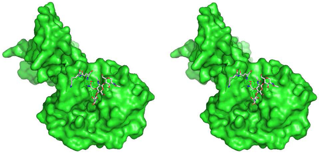
The catalytic module of the Cpl-1 endolysin in complex with peptidoglycan (PDB Code 2J8G). A comparison of this protein structure with those of Figs. 2 and 4 emphasizes the evolutionary convergence of different protein motifs for the purpose of peptidoglycan binding.
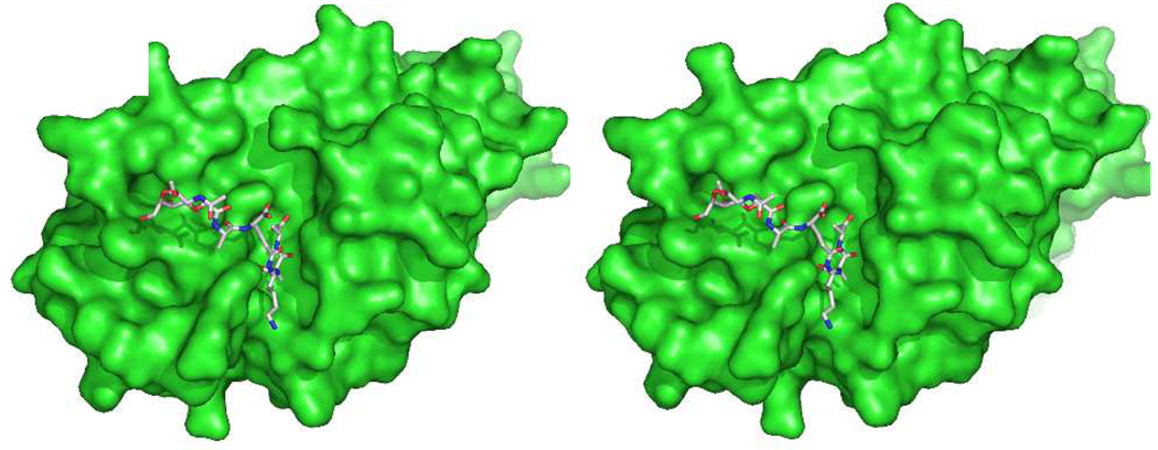
Complex of E. coli PBP6 (catalytic domain of monomer B, PDB Code 3ITB) with the peptidoglycan structure that is shown in Scheme 7.
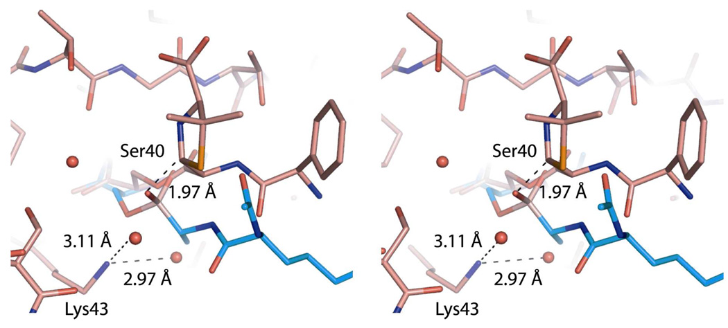
Stereo view of the PBP6 acyl-enzyme complex compared to that of the deacylation transition state. A PBP5 transition-state analog structure (PDB Code 1Z6F) is superimposed onto the PBP6 structure with ampicillin (PDB Code 3ITA), showing active site residues 39, 40, 43, 108 and 210. The transition-state analog and the catalytic serine of PBP5 are shown in blue, whereas ampicillin and PBP6 are shown in pink. Water molecules are represented as red spheres. Key distances are displayed as dashed lines. The amine group of Lys43 engages in hydrogen bonds with two water molecules in the PBP6 acyl-enzyme complex. The distance between the β-lactam-thiazolidine bridging carbon atom of ampicillin, and the boronic acid oxygen of the superimposed transition state analog, is only 1.97 Å. This carbon presents a steric impediment to hydrolytic deacylation.,
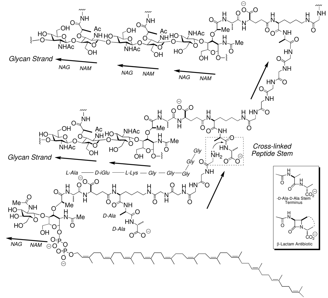
The cross-linking event in peptidoglycan biosynthesis by the Gram-positive bacterium S. aureus. The structure of the immediate biosynthetic precursor for the peptidoglycan of S. aureus, Lipid II-Gly5 is shown as the bottom structure. The cross-linking event is highlighted within the hashed-edged box shown in the center right. This stem cross-linking event, wherein the amine terminus of the pentaaglycine displaces the d -alanine terminus on the stem of an adjacent glycan strand, is catalyzed by the transpeptidase domain of a high molecular mass PBP enzyme. This reaction proceeds through an acyl-enzyme intermediate (not depicted). Lipid II-Gly5 participation in transpeptidase cross-linking is shown only for the purpose of illustration. Peptide stem cross-linking almost certainly occurs subsequent to Lipid II-Gly5-dependent, transglycosylase-catalyzed elongation of the glycan strand. The inset box (lower right) shows the β-lactam antibiotic structure as a mimetic of a conformation of the -d -Ala-d -Ala stem used in the cross-linking.
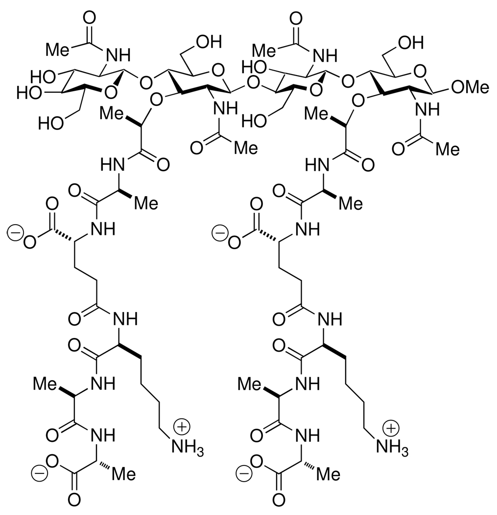
The synthetic [NAG-NAM]2 mimetic of the non-crosslinked peptidoglycan of the cell wall, used for the NMR determination of the solution conformation of a peptidoglycan strand.
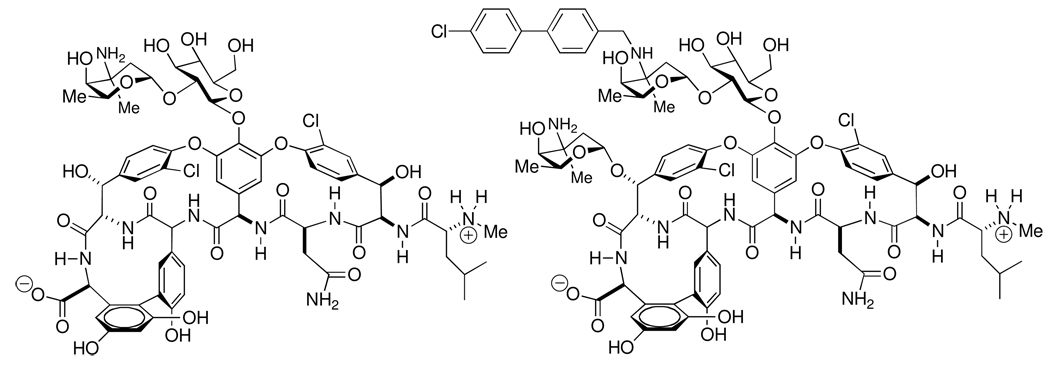
Structures of vancomycin (left) and oritavancin (right).
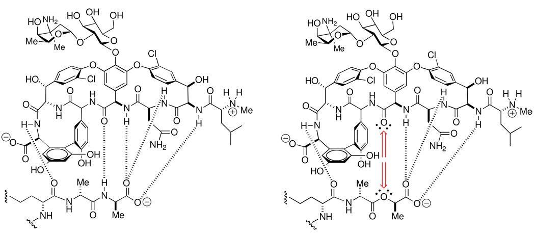
Hydrogen-bonding interactions in the -d -Ala-d -Ala peptidoglycan-vancomycin host-guest complex (left) compared to the -d -Ala-d -Lac peptidoglycan-vancomycin host-guest complex (right). The loss of a key hydrogen bond, and its replacement by unfavorable non-bonding repulsive interactions, is indicated by the red-colored arrows in the -d -Ala-d -Lac depsipeptide complex.
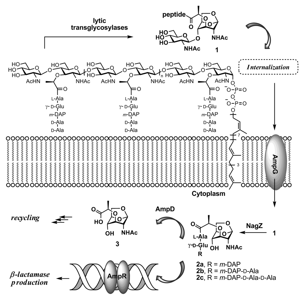
Peptidoglycan fragmentation leads to NAG-NAM segments, which are transported across plasma membrane to initiate the recycling of these segments and gene induction for production of the β-lactamase enzyme.
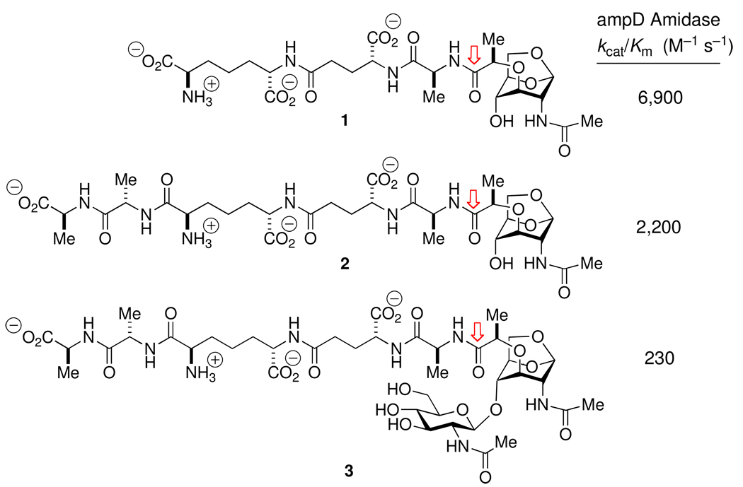
Substrate specificity, measured by kcat/Km, for Citrobacter freundii AmpD amidase-catalyzed amide bond hydrolysis (cleavage point indicated by the arrows) of three anhydromuramyl muropeptide structures.
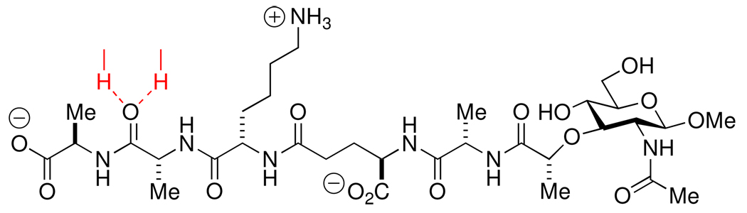
Structure of the synthetic peptidoglycan, used in the determination of the crystal structure of a Michaelis complex-like structure with E. coli PBP6. The red-colored hydrogen bonds drawn to the (scissile) amide carbonyl of the penultimate d -Ala residue indicates its occupancy of the oxyanion hole of the active site.
Similar articles
-
Modifications to the peptidoglycan backbone help bacteria to establish infection.Infect Immun. 2011 Feb;79(2):562-70. doi: 10.1128/IAI.00651-10. Epub 2010 Nov 1. Infect Immun. 2011. PMID: 21041496 Free PMC article. Review.
-
The medium is the message: interspecies and interkingdom signaling by peptidoglycan and related bacterial glycans.Annu Rev Microbiol. 2014;68:137-54. doi: 10.1146/annurev-micro-091213-112844. Epub 2014 May 16. Annu Rev Microbiol. 2014. PMID: 24847956 Review.
-
Resistance to peptidoglycan-degrading enzymes.Crit Rev Microbiol. 2020 Nov;46(6):703-726. doi: 10.1080/1040841X.2020.1825333. Epub 2020 Sep 26. Crit Rev Microbiol. 2020. PMID: 32985279 Review.
-
Natural Products at Work: Structural Insights into Inhibition of the Bacterial Membrane Protein MraY.Angew Chem Int Ed Engl. 2016 Sep 19;55(39):11722-4. doi: 10.1002/anie.201606396. Epub 2016 Aug 11. Angew Chem Int Ed Engl. 2016. PMID: 27511599
-
Building peptidoglycan inside eukaryotic cells: A view from symbiotic and pathogenic bacteria.Mol Microbiol. 2020 Mar;113(3):613-626. doi: 10.1111/mmi.14452. Mol Microbiol. 2020. PMID: 32185832 Free PMC article. Review.
Cited by
-
Antibacterial Properties of Graphene-Based Nanomaterials.Nanomaterials (Basel). 2019 May 13;9(5):737. doi: 10.3390/nano9050737. Nanomaterials (Basel). 2019. PMID: 31086043 Free PMC article. Review.
-
Structure and cell wall cleavage by modular lytic transglycosylase MltC of Escherichia coli.ACS Chem Biol. 2014 Sep 19;9(9):2058-66. doi: 10.1021/cb500439c. Epub 2014 Jul 10. ACS Chem Biol. 2014. PMID: 24988330 Free PMC article.
-
Early insights into the interactions of different β-lactam antibiotics and β-lactamase inhibitors against soluble forms of Acinetobacter baumannii PBP1a and Acinetobacter sp. PBP3.Antimicrob Agents Chemother. 2012 Nov;56(11):5687-92. doi: 10.1128/AAC.01027-12. Epub 2012 Aug 20. Antimicrob Agents Chemother. 2012. PMID: 22908165 Free PMC article.
-
Identification of products of inhibition of GES-2 beta-lactamase by tazobactam by x-ray crystallography and spectrometry.J Biol Chem. 2011 Apr 22;286(16):14396-409. doi: 10.1074/jbc.M110.208744. Epub 2011 Feb 22. J Biol Chem. 2011. PMID: 21345789 Free PMC article.
-
Messenger functions of the bacterial cell wall-derived muropeptides.Biochemistry. 2012 Apr 10;51(14):2974-90. doi: 10.1021/bi300174x. Epub 2012 Mar 27. Biochemistry. 2012. PMID: 22409164 Free PMC article. Review.
References
-
- Coyette J, van der Ende A. Peptidoglycan: the bacterial Achilles heel. FEMS Microbiol. Rev. 2008;32:147–148. - PubMed
-
- Schneider T, Sahl H-G. An oldie but a goodie - cell wall biosynthesis as antibiotic target pathway. Int. J. Med. Microbiol. 2010;300:161–169. - PubMed
-
- Daniel RA, Errington J. Control of cell morphogenesis in bacteria: two distinct ways to make a rod-shaped cell. Cell. 2003;113:767–776. - PubMed
Publication types
MeSH terms
Substances
Grants and funding
LinkOut - more resources
Full Text Sources
Medical
