

Patterns of Expression of Vaginal T-Cell Activation Markers during Estrogen-Maintained Vaginal Candidiasis
- PMID: 20525139
- PMCID: PMC2868890
- DOI: 10.1186/1710-1492-4-4-157
Patterns of Expression of Vaginal T-Cell Activation Markers during Estrogen-Maintained Vaginal Candidiasis
Abstract
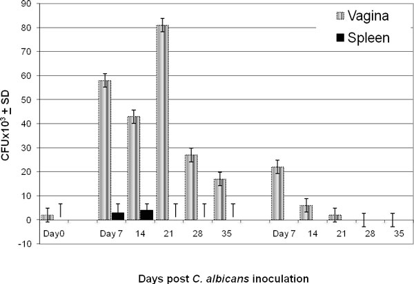
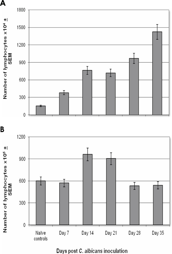
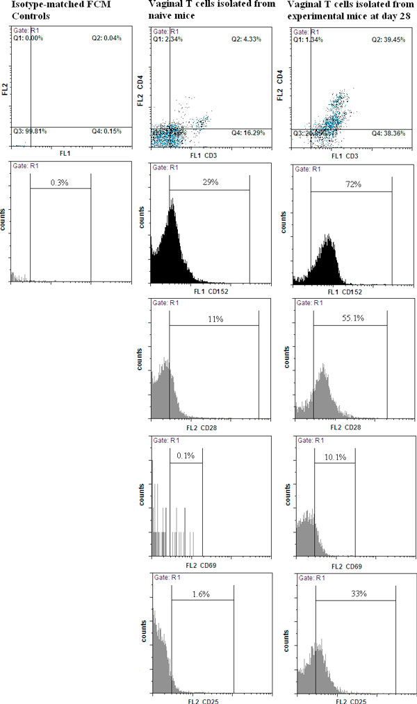
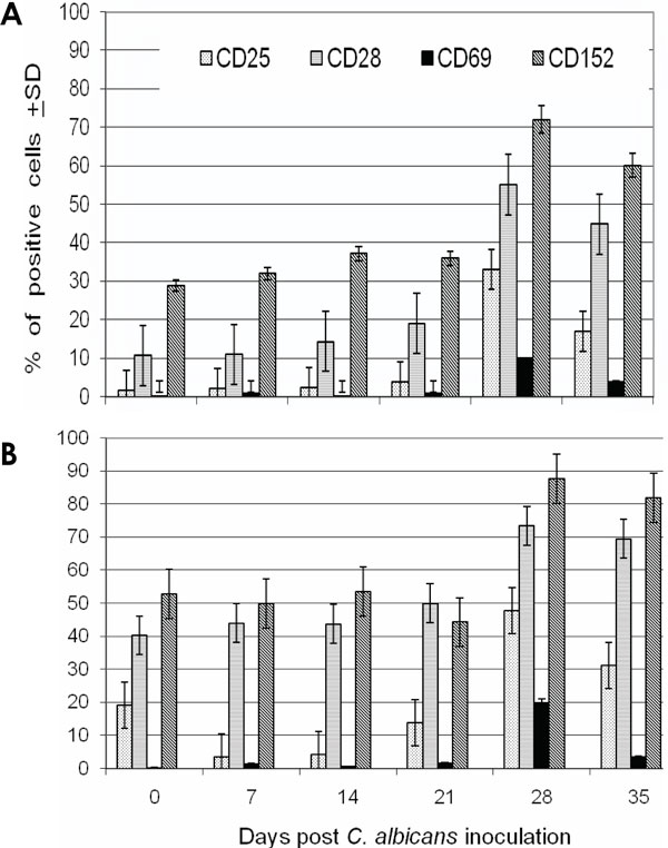
: The immunosuppressive activity of estrogen was further investigated by assessing the pattern of expression of CD25, CD28, CD69, and CD152 on vaginal T cells during estrogen-maintained vaginal candidiasis. A precipitous and significant decrease in vaginal fungal burden toward the end of week 3 postinfection was concurrent with a significant increase in vaginal lymphocyte numbers. During this period, the percentage of CD3+, CD3+CD4+, CD152+, and CD28+ vaginal T cells gradually and significantly increased. The percentage of CD3+ and CD3+CD4+ cells increased from 43% and 15% at day 0 to 77% and 40% at day 28 postinfection. Compared with 29% CD152+ vaginal T cells in naive mice, > 70% of vaginal T cells were CD152+ at day 28 postinfection. In conclusion, estrogen-maintained vaginal candidiasis results in postinfection time-dependent changes in the pattern of expression of CD152, CD28, and other T-cell markers, suggesting that T cells are subject to mixed suppression and activation signals.
Figures
Similar articles
-
Alterations of the expression of T-cell-related costimulatory CD28 and downregulatory CD152 (CTLA-4) molecules in patients with B-cell chronic lymphocytic leukaemia.Br J Cancer. 2004 May 17;90(10):2042-8. doi: 10.1038/sj.bjc.6601833. Br J Cancer. 2004. PMID: 15138491 Free PMC article.
-
Expression of the costimulator molecules, CD80, CD86, CD28, and CD152 on lymphocytes from neonates and young children.Hum Immunol. 1999 Nov;60(11):1039-48. doi: 10.1016/s0198-8859(99)00090-7. Hum Immunol. 1999. PMID: 10600000
-
Cyclic adenosine 5'-monophosphate and calcium induce CD152 (CTLA-4) up-regulation in resting CD4+ T lymphocytes.J Immunol. 2002 Dec 1;169(11):6231-5. doi: 10.4049/jimmunol.169.11.6231. J Immunol. 2002. PMID: 12444128
-
Surface CD152 (CTLA-4) expression and signaling dictates longevity of CD28null T cells.J Immunol. 2009 May 1;182(9):5342-51. doi: 10.4049/jimmunol.0801624. J Immunol. 2009. PMID: 19380781
-
Abnormal T-cell function in B-cell chronic lymphocytic leukaemia.Leuk Lymphoma. 2003 Mar;44(3):383-9. doi: 10.1080/1042819021000029993. Leuk Lymphoma. 2003. PMID: 12688308 Review.
Cited by
-
Tissue-resident memory T cells.Immunol Rev. 2013 Sep;255(1):165-81. doi: 10.1111/imr.12087. Immunol Rev. 2013. PMID: 23947354 Free PMC article. Review.
-
Estrogen treatment predisposes to severe and persistent vaginal candidiasis in diabetic mice.J Diabetes Metab Disord. 2014 Jan 8;13(1):15. doi: 10.1186/2251-6581-13-15. J Diabetes Metab Disord. 2014. PMID: 24401317 Free PMC article.
References
LinkOut - more resources
Full Text Sources
Research Materials
