FRET microscopy autologous tumor lysate processing in mature dendritic cell vaccine therapy
- PMID: 20525246
- PMCID: PMC2894751
- DOI: 10.1186/1479-5876-8-52
FRET microscopy autologous tumor lysate processing in mature dendritic cell vaccine therapy
Abstract
Background: Antigen processing by dendritic cells (DC) exposed to specific stimuli has been well characterized in biological studies. Nonetheless, the question of whether autologous whole tumor lysates (as used in clinical trials) are similarly processed by these cells has not yet been resolved.
Methods: In this study, we examined the transfer of peptides from whole tumor lysates to major histocompatibility complex class II molecules (MHC II) in mature dendritic cells (mDC) from a patient with advanced melanoma. Tumor antigenic peptides-MHC II proximity was revealed by Förster Resonance Energy Transfer (FRET) measurements, which effectively extends the application of fluorescence microscopy to the molecular level (<100A). Tumor lysates were labelled with Alexa-488, as the donor, and mDC MHC II HLA-DR molecules were labelled with Alexa-546-conjugated IgG, as the acceptor.
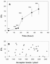
Results: We detected significant energy transfer between donor and acceptor-labelled antibodies against HLA-DR at the membrane surface of mDC. FRET data indicated that fluorescent peptide-loaded MHC II molecules start to accumulate on mDC membranes at 16 hr from the maturation stimulus, steeply increasing at 22 hr with sustained higher FRET detected up to 46 hr.
Conclusions: The results obtained imply that the patient mDC correctly processed the tumor specific antigens and their display on the mDC surface may be effective for several days. These observations support the rationale for immunogenic efficacy of autologous tumor lysates.
Figures


Similar articles
-
Tumor cell lysates as immunogenic sources for cancer vaccine design.Hum Vaccin Immunother. 2014;10(11):3261-9. doi: 10.4161/21645515.2014.982996. Hum Vaccin Immunother. 2014. PMID: 25625929 Free PMC article. Review.
-
[Anti-lung cancer effect of myeloid and plasmacytoid dendritic cell combined vaccines loaded with tumor cell lysates in vitro].Zhonghua Zhong Liu Za Zhi. 2019 Jul 23;41(7):501-507. doi: 10.3760/cma.j.issn.0253-3766.2019.07.004. Zhonghua Zhong Liu Za Zhi. 2019. PMID: 31357836 Chinese.
-
Binding of prostate-specific membrane antigen to dendritic cells: a critical step in vaccine preparation.Cytotherapy. 2009;11(8):1090-100. doi: 10.3109/14653240903164971. Cytotherapy. 2009. PMID: 19929472
-
Exosomes as potent cell-free peptide-based vaccine. I. Dendritic cell-derived exosomes transfer functional MHC class I/peptide complexes to dendritic cells.J Immunol. 2004 Feb 15;172(4):2126-36. doi: 10.4049/jimmunol.172.4.2126. J Immunol. 2004. PMID: 14764678
-
Dendritic cell gene therapy.Surg Oncol Clin N Am. 2002 Jul;11(3):645-60. doi: 10.1016/s1055-3207(02)00027-3. Surg Oncol Clin N Am. 2002. PMID: 12487060 Review.
Cited by
-
Vaccination with autologous dendritic cells loaded with autologous tumor lysate or homogenate combined with immunomodulating radiotherapy and/or preleukapheresis IFN-α in patients with metastatic melanoma: a randomised "proof-of-principle" phase II study.J Transl Med. 2014 Jul 22;12:209. doi: 10.1186/1479-5876-12-209. J Transl Med. 2014. PMID: 25053129 Free PMC article. Clinical Trial.
-
Vaccination with dendritic cells loaded with tumor apoptotic bodies (Apo-DC) in patients with chronic lymphocytic leukemia: effects of various adjuvants and definition of immune response criteria.Cancer Immunol Immunother. 2012 Jun;61(6):865-79. doi: 10.1007/s00262-011-1149-5. Epub 2011 Nov 16. Cancer Immunol Immunother. 2012. PMID: 22086161 Free PMC article.
-
Immunity to melanin and to tyrosinase in melanoma patients, and in people with vitiligo.BMC Complement Altern Med. 2012 Jul 26;12:109. doi: 10.1186/1472-6882-12-109. BMC Complement Altern Med. 2012. PMID: 22834951 Free PMC article.
-
In vitro transfection of bone marrow-derived dendritic cells with TATp-liposomes.Int J Nanomedicine. 2014 Feb 13;9:963-73. doi: 10.2147/IJN.S53432. eCollection 2014. Int J Nanomedicine. 2014. PMID: 24611012 Free PMC article.
References
-
- Hart DN. Dendritic cells: unique leukocyte populations which control the primary immune response. Blood. 1997;90:3245–3287. - PubMed
-
- Banchereau J, Palucka AK, Dhodapkar M, Burkeholder S, Taquet N, Rolland A, Taquet S, Coquery S, Wittkowski KM, Bhardwaj N, Pineiro L, Steinman R, Fay J. Immune and clinical responses in patients with metastatic melanoma to CD34(+) progenitor-derived dendritic cell vaccine. Cancer Res. 2001;61:6451–6458. - PubMed
Publication types
MeSH terms
Substances
LinkOut - more resources
Full Text Sources
Research Materials

