

Immunophenotypic features of tumor infiltrating lymphocytes from mammary carcinomas in female dogs associated with prognostic factors and survival rates
- PMID: 20525350
- PMCID: PMC2894795
- DOI: 10.1186/1471-2407-10-256
Immunophenotypic features of tumor infiltrating lymphocytes from mammary carcinomas in female dogs associated with prognostic factors and survival rates
Abstract
Background: The immune system plays an important role in the multifactorial biologic system during the development of neoplasias. However, the involvement of the inflammatory response in the promotion/control of malignant cells is still controversial, and the cell subsets and the mechanisms involved are poorly investigated. The goal of this study was to characterize the clinical-pathological status and the immunophenotyping profile of tumor infiltrating lymphocytes and their association with the animal survival rates in canine mammary carcinomas.
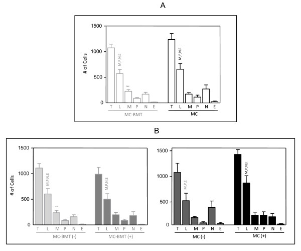
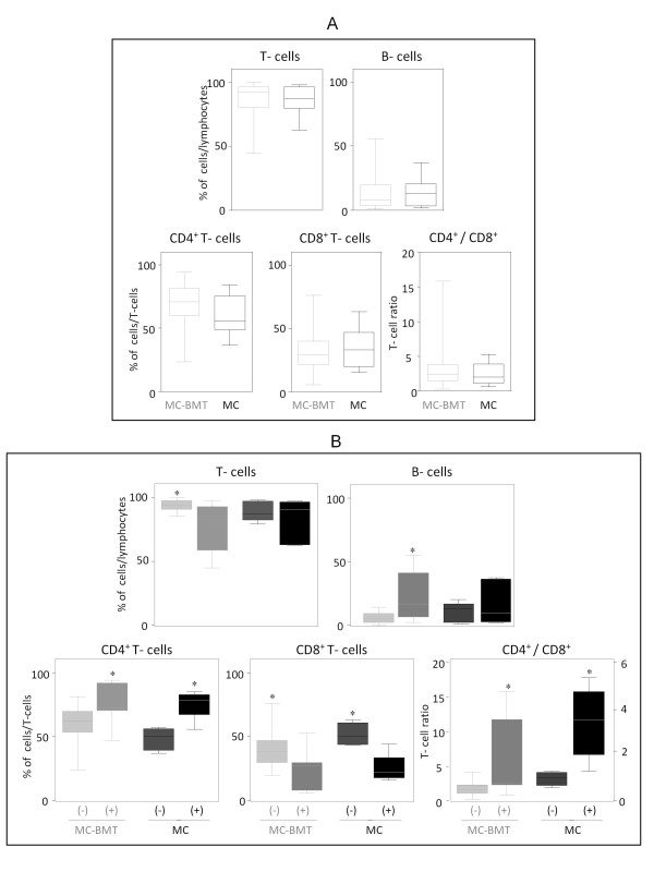
Methods: Fifty-one animals with mammary carcinomas, classified as carcinomas in mixed tumors-MC-BMT = 31 and carcinomas-MC = 20 were submitted to systematic clinical-pathological analysis (tumor size; presence of lymph node and pulmonary metastasis; clinical stage; histological grade; inflammatory distribution and intensity as well as the lymphocytic infiltrate intensity) and survival rates. Twenty-four animals (MC-BMT = 16 and MC = 8) were elected to the immunophenotypic study performed by flow cytometry.
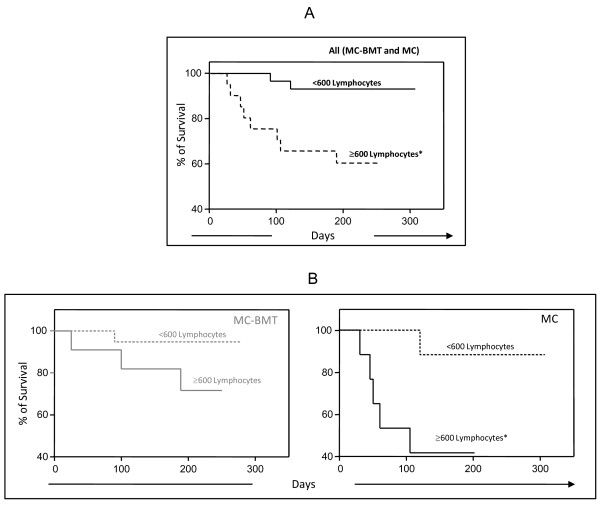
Results: Data analysis demonstrated that clinical stage II-IV and histological grade was I more frequent in MC-BMT as compared to MC. Univariate analysis demonstrated that the intensity of inflammation (moderate/intense) and the proportion of CD4+ (> or = 66.7%) or CD8+ T-cells (<33.3%) were not associated with worse survival rate. Multivariate analysis demonstrated that only lymphocytic infiltrate intensity > or = 600 (P = 0.02) remained as independent prognostic factor. Despite the clinical manifestation, the lymphocytes represented the predominant cell type in the tumor infiltrate. The percentage of T-cells was higher in animals with MC-BMT without metastasis, while the percentage of B-lymphocytes was greater in animals with metastasized MC-BMT (P < 0.05). The relative percentage of CD4+ T-cells was significantly greater in metastasized tumors (both MC-BMT and MC), (P < 0.05) while the proportion of CD8+ T-cells was higher in MC-BMT without metastasis. Consequently, the CD4+/CD8+ ratio was significantly increased in both groups with metastasis. Regardless of the tumor type, the animals with high proportions of CD4+ and low CD8+ T-cells had decreased survival rates.
Conclusion: The intensity of lymphocytic infiltrate and probably the relative abundance of the CD4+ and CD8+ T-lymphocytes may represent important survival prognostic biomarkers for canine mammary carcinomas.
Figures


References
-
- Misdorp W. In: Tumors in Domestic Animals. 4. Meuten DJ, editor. Iowa State: University of California; 2002. Tumors of the mammary gland; pp. 575–606. full_text.
-
- Peleteiro MC. Tumores mamários na cadela e na gata. Revista Portuguesa de Ciências Veterinárias. 1994;89(509):10–29.
-
- Cassali GD, Gobbi H, Malm C, Schmitt FC. Evaluation of accuracy of fine needle aspiration cytology for diagnosis of canine mammary tumours: comparative features with human tumours. Cytopathology. 2007;18(3):191–6. - PubMed
-
- Bertagnolli AC, Cassali GD, Genelhu MCLS, Costa FA, Oliveira JFC, Gonçalves PBD. Immunohistochemical expression of p63 and {Delta}Np63 in mixed tumors of canine mammary glands and its relation with p53 expression. Veterinary Pathology. 2009;46:407–415. doi: 10.1354/vp.08-VP-0128-C-FL. - DOI - PubMed
Publication types
MeSH terms
LinkOut - more resources
Full Text Sources
Research Materials
