. 2010 Aug 6;285(32):24494-507.
doi: 10.1074/jbc.M110.130575. Epub 2010 Jun 4.

Essential in vivo roles of the C-type lectin receptor CLEC-2: embryonic/neonatal lethality of CLEC-2-deficient mice by blood/lymphatic misconnections and impaired thrombus formation of CLEC-2-deficient platelets

Affiliations

Essential in vivo roles of the C-type lectin receptor CLEC-2: embryonic/neonatal lethality of CLEC-2-deficient mice by blood/lymphatic misconnections and impaired thrombus formation of CLEC-2-deficient platelets

Katsue Suzuki-Inoue et al. J Biol Chem. .

Abstract

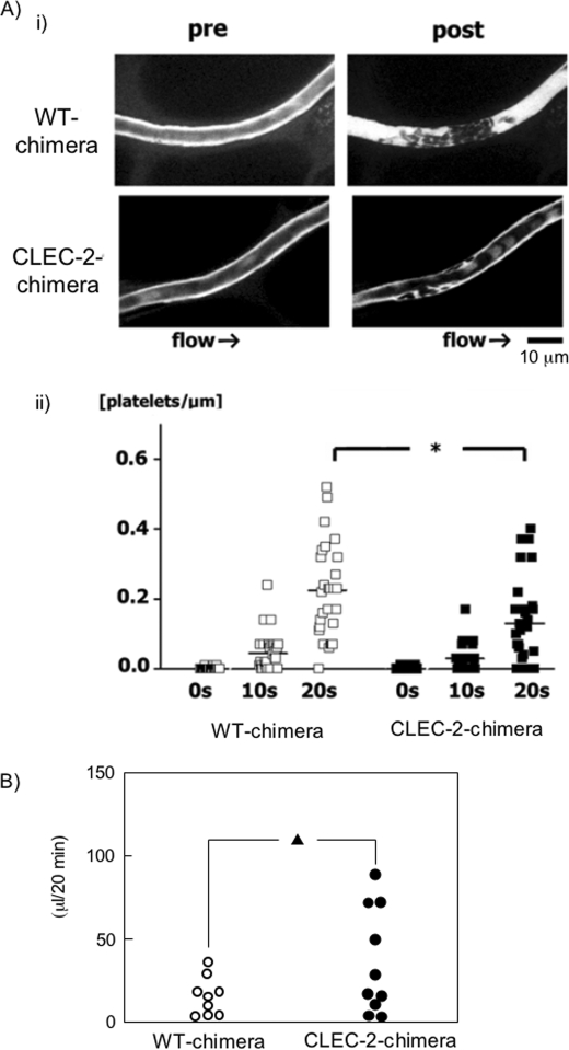
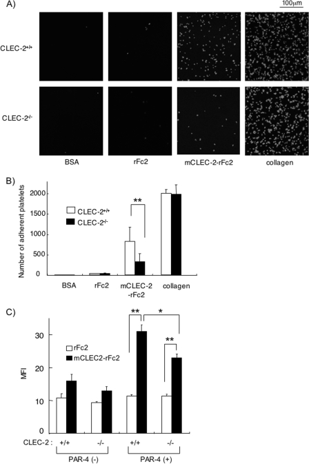
CLEC-2 has been described recently as playing crucial roles in thrombosis/hemostasis, tumor metastasis, and lymphangiogenesis. The snake venom rhodocytin is known as a strong platelet activator, and we have shown that this effect is mediated by CLEC-2 (Suzuki-Inoue, K., Fuller, G. L., García, A., Eble, J. A., Pöhlmann, S., Inoue, O., Gartner, T. K., Hughan, S. C., Pearce, A. C., Laing, G. D., Theakston, R. D., Schweighoffer, E., Zitzmann, N., Morita, T., Tybulewicz, V. L., Ozaki, Y., and Watson, S. P. (2006) Blood 107, 542-549). Podoplanin, which is expressed on the surface of tumor cells, is an endogenous ligand for CLEC-2 and facilitates tumor metastasis by inducing platelet aggregation. Mice deficient in podoplanin, which is also expressed on the surface of lymphatic endothelial cells, show abnormal patterns of lymphatic vessel formation. In this study, we report on the generation and phenotype of CLEC-2-deficient mice. These mice are lethal at the embryonic/neonatal stages associated with disorganized and blood-filled lymphatic vessels and severe edema. Moreover, by transplantation of fetal liver cells from Clec-2(-/-) or Clec-2(+/+) embryos, we were able to demonstrate that CLEC-2 is involved in thrombus stabilization in vitro and in vivo, possibly through homophilic interactions without apparent increase in bleeding tendency. We propose that CLEC-2 could be an ideal novel target protein for an anti-platelet drug, which inhibits pathological thrombus formation but not physiological hemostasis.


Figures

FIGURE 1.
FIGURE 1.
Lymphatic function is impaired in Clec-2−/− embryos. A, lateral views of E15.5 Clec-2−/− (left) and Clec-2+/+ (right) embryos. Back edema in the Clec-2−/− embryo is indicated by the arrow. B, lymphangiography by injection of FITC-dextran into the forelimbs of Clec-2+/+ (upper panel) and Clec-2−/− (lower panel) embryos at E17.5. Injection sites on the forelimbs are indicated by the arrows. The arrowheads indicate visualized collecting lymphatic vessels in the Clec-2+/+ embryo. The schematics on the right illustrate the visualized collecting lymphatic vessel in Clec-2+/+ mice (upper panel) and injection sites in Clec-2+/+ (upper panel) and Clec-2−/− (lower panel) embryos (arrow). Rt, right.
FIGURE 2.
FIGURE 2.
Developing lymphatic circulation in mice lacking CLEC-2 communicates with the blood circulation. A, mesenteric sections of E15.5 Clec-2+/+ (upper panels) and Clec-2−/− (lower panels) embryos stained with hematoxylin and eosin (HE; left panels) and LYVE-1 (right panels). B, confocal images of intestinal cryosections of E17.5 Clec-2+/+ (upper panels) and Clec-2−/− (lower panels) embryos with antibodies against PECAM-1, α-smooth muscle actin (α-SMA), and LYVE-1. L, lymphatic vessels; V, vein; A, arteries. The arrows indicate blood cells.
FIGURE 3.
FIGURE 3.
Blood-filled disorganized lymphatic vessels and abnormal connection between blood and lymphatic vessels in Clec-2−/− embryos. Whole-mount triple fluorescence confocal microscopy of embryonic back skin was performed with antibodies to PECAM-1 (red), LYVE-1 (green), and TER-119 (blue) at E14.5 (A–I) and E17.5 (J–L). A–F, whereas blood vessels visualized by PECAM-1 staining appear unaffected in Clec-2−/− embryos, lymphatic vessels visualized by LYVE-1 staining are disorganized and distended in Clec-2−/− embryos. Lymphatic vessels are filled with TER-119+ erythrocytes (arrows) in Clec-2−/− embryos. G–L, abnormal connection sites (arrowheads) between blood and lymphatic vessels were detected in Clec-2−/− embryos. Scale bars = 100 μm.
FIGURE 4.
FIGURE 4.
Responses to rhodocytin are abolished in CLEC-2-deficient platelets, but WT platelets respond normally to other agonists. A, shown is a Western blot (WB) of washed platelets from two WT chimeras and two CLEC-2 chimeras with anti-mouse CLEC-2 antibody. The arrow indicates murine CLEC-2 (mCLEC-2). B, whole blood from WT and CLEC-2 chimeras was diluted 15-fold with modified Tyrode's buffer. 25 μl of the diluted whole blood was incubated with Cy2-conjugated anti-mouse CLEC-2 antibody or Cy2-conjugated control rabbit IgG for 15 min at room temperature. Reactions were terminated by the addition of 400 μl of PBS, and the samples were then analyzed using a FACScan. C, washed platelets from WT or CLEC-2 chimeras were used for aggregation studies. The washed platelets were stimulated by the indicated agonists, and platelet aggregation was monitored by light transmission using a Born aggregometer at 37 °C for 10 min. D, the activation of integrin αIIbβ3 induced by the indicated agonists was investigated. Whole blood from WT or CLEC-2 chimeras was diluted 15-fold with modified Tyrode's buffer. 25 μl of diluted whole blood was stimulated with the indicated platelet agonists for 5 min at room temperature, followed by the addition of FITC-conjugated control rat IgG or FITC-conjugated anti-activated mouse integrin αIIbβ3 (clone Jon-A) for 15 min at room temperature. Reactions were terminated by the addition of 400 μl of PBS, and the samples were then analyzed using a FACScan. Data are expressed as the mean of the median fluorescence intensity (MFI) S.E. (n = 4–7). E, CD62P expression stimulated by the indicated agonists was investigated. 25 μl of washed platelets (5 × 10⁷/ml) was stimulated with the indicated platelet agonists for 5 min at room temperature, followed by the addition of PE-conjugated control rat IgG or PE-conjugated anti-mouse CD62P for 15 min at room temperature. Reactions were terminated by the addition of 400 μl of PBS, and the samples were then analyzed using a FACScan. Data are expressed as the mean of the median fluorescence intensity ± S.E. (n = 4–7). F, serotonin release from dense granules was investigated. Washed platelets (3 × 10⁸/ml) were stimulated with the indicated platelet agonists for 5 min. After platelets were removed by centrifugation, the serotonin concentration of the supernatant was measured by enzyme-linked immunosorbent assay. Serotonin release is expressed as the percent serotonin concentration of the platelet lysate. Data are expressed as the mean ± S.E. (n = 3). *, p < 0.05; **, p < 0.005. rhod, rhodocytin; col, collagen; rest, resting.
FIGURE 5.
FIGURE 5.
CLEC-2-deficient platelets showed normal adhesion and spreading on the surface of collagen, fibrinogen, laminin, and vWF. A, platelet spreading on the surface of major extracellular matrices was investigated. Washed platelets from WT chimeras (Clec-2+/+) or CLEC-2 chimeras (Clec-2−/−) were seeded on coverslips coated with laminin (LN), collagen (Col), fibrinogen (Fib), or vWF for 30 min at room temperature in the presence or absence of 10 μm ADP. Adherent platelets were fixed in 3% paraformaldehyde, permeabilized with 0.3% Triton X-100 for 5 min, and stained with TRITC-conjugated phalloidin. Platelets were visualized using an inverted fluorescence microscope and a digital camera. B, shown is the quantification of adherent platelets in the images in A. BSA-coated coverslips were prepared as a negative control. At least six images from two independent experiments were chosen at random per experiment and analyzed by two individuals, one of whom performed the analysis under blind conditions. Adherent platelets were counted (0.006 mm2/image), and platelet surface area was analyzed using NIH Image.
FIGURE 6.
FIGURE 6.
Thrombus formation on the surface of collagen under flow conditions is impaired in CLEC-2 chimeras. A, shown are video stills of thrombus formation on the surface of collagen under flow conditions. Capillary tubes were coated with 50 μg/ml collagen and blocked with PBS containing 2% BSA. Whole blood from WT chimeras (Clec-2+/+) or CLEC-2 chimeras (Clec-2−/−) anticoagulated with PPACK and heparin that had been pretreated with 3,3′-dihexyloxacarbocyanine iodide was perfused into capillaries at 2000 s−1, and adherent platelets were visualized using a fluorescence video microscope. Movie data were converted into sequential photo images. B, shown are three-dimensional images of the thrombus formation. After perfusion of the blood, capillaries with thrombus were visualized using an Olympus FV-1000 confocal microscope. C, for measurement of thrombus volume, the images were analyzed using FluoView software, and the relative thrombus volume is expressed as integrated fluorescence intensity (IFI). *, p < 0.05 (n = at least 5 from two different mice).
FIGURE 7.
FIGURE 7.
In vivo thrombus formation in a laser-induced injury model is impaired in CLEC-2 chimeras without significant increase in tail bleeding. A, video stills of mesenteric capillaries were obtained by intravital fluorescence microscopy before (pre) and 20 s after (post) laser-induced injury (panel i). The numbers of platelets in developing thrombi after laser injury to capillaries were calculated (panel ii). The y axis represents the numbers of platelets/micrometer of obtained vessel length. Results from WT and CLEC-2 chimeras 7 weeks after transplantation (17 weeks old) are shown (n = 5 each). *, p < 0.05. B, shown is the tail bleeding in WT and CLEC-2 chimeras. Each symbol represents one individual. Results from WT and CLEC-2 chimeras 8 weeks after transplantation (17 weeks old) are shown. ▴, not significant (p = 0.08).
FIGURE 8.
FIGURE 8.
CLEC-2 forms a homophilic association. Different concentrations of hCLEC-2-rFc2 (A) or mCLEC-2-rFc2 (B) were flowed over an immobilized hCLEC-2-rFc2 (A), mCLEC-2-rFc2 (B), or control surface coated with rFc2. The arrows indicate the beginning and end of perfusion of hCLEC-2-rFc2 and mCLEC-2-rFc2. The results are shown from one experiment that is representative of four others. RU, resonance units. Kd ± S.E. (n = 4) of homophilic association of hCLEC-2-rFc2 or mCLEC-2-rFc2 was determined as described under “Experimental Procedures” (C).
FIGURE 9.
FIGURE 9.
Platelet adhesion to mCLEC-2-rFc2-coated surfaces and soluble mCLEC-2-rFc2 binding to platelets are inhibited in CLEC-2-deficient platelets. A, shown is platelet adhesion on a surface coated with mCLEC-2-rFc2. Washed murine platelets from WT or CLEC-2 chimeras were seeded on coverslips coated with the indicated materials. Adherent platelets were fixed in paraformaldehyde, permeabilized with 0.3% Triton X-100, and stained with TRITC-conjugated phalloidin for 2 h. Platelets were visualized using an inverted fluorescence microscope and a digital camera. B, shown is the quantification of the platelet adhesion in A. At least six images from two independent experiments were chosen at random per experiment and analyzed by two individuals, one of whom performed the analysis under blind conditions. Adherent platelets were counted (0.006 mm2/image). C, the binding of soluble mCLEC-2-rFc2 or rFc2 on the surface of WT (Clec-2+/+) or CLEC-2-deficient (Clec-2−/−) platelets was investigated by flow cytometry. Quantification of the soluble protein binding was performed using median fluorescence intensity (MFI). Data are expressed as mean ± S.E. (n > 4). Statistical significance was evaluated by Student's t test. In each case, p values <0.05 were taken as the minimum to indicate statistical significance. *, p < 0.05; **, p < 0.005.

Similar articles

Cited by

References

    1. Cambi A., Figdor C. G. (2003) Curr. Opin. Cell Biol. 15, 539–546 - PubMed
    1. Marshall A. S., Gordon S. (2004) Eur. J. Immunol. 34, 18–24 - PubMed
    1. Suzuki-Inoue K., Fuller G. L., García A., Eble J. A., Pöhlmann S., Inoue O., Gartner T. K., Hughan S. C., Pearce A. C., Laing G. D., Theakston R. D., Schweighoffer E., Zitzmann N., Morita T., Tybulewicz V. L., Ozaki Y., Watson S. P. (2006) Blood 107, 542–549 - PubMed
    1. Suzuki-Inoue K., Kato Y., Inoue O., Kaneko M. K., Mishima K., Yatomi Y., Yamazaki Y., Narimatsu H., Ozaki Y. (2007) J. Biol. Chem. 282, 25993–26001 - PubMed
    1. Christou C. M., Pearce A. C., Watson A. A., Mistry A. R., Pollitt A. Y., Fenton-May A. E., Johnson L. A., Jackson D. G., Watson S. P., O'Callaghan C. A. (2008) Biochem. J. 411, 133–140 - PMC - PubMed

Publication types