. 2010 Aug 13;285(33):25269-84.
doi: 10.1074/jbc.M110.144311. Epub 2010 Jun 4.

YopD self-assembly and binding to LcrV facilitate type III secretion activity by Yersinia pseudotuberculosis

Affiliations

YopD self-assembly and binding to LcrV facilitate type III secretion activity by Yersinia pseudotuberculosis

Tiago R D Costa et al. J Biol Chem. .

Abstract

YopD-like translocator proteins encoded by several Gram-negative bacteria are important for type III secretion-dependent delivery of anti-host effectors into eukaryotic cells. This probably depends on their ability to form pores in the infected cell plasma membrane, through which effectors may gain access to the cell interior. In addition, Yersinia YopD is a negative regulator essential for the control of effector synthesis and secretion. As a prerequisite for this functional duality, YopD may need to establish molecular interactions with other key T3S components. A putative coiled-coil domain and an alpha-helical amphipathic domain, both situated in the YopD C terminus, may represent key protein-protein interaction domains. Therefore, residues within the YopD C terminus were systematically mutagenized. All 68 mutant bacteria were first screened in a variety of assays designed to identify individual residues essential for YopD function, possibly by providing the interaction interface for the docking of other T3S proteins. Mirroring the effect of a full-length yopD gene deletion, five mutant bacteria were defective for both yop regulatory control and effector delivery. Interestingly, all mutations clustered to hydrophobic amino acids of the amphipathic domain. Also situated within this domain, two additional mutants rendered YopD primarily defective in the control of Yop synthesis and secretion. Significantly, protein-protein interaction studies revealed that functionally compromised YopD variants were also defective in self-oligomerization and in the ability to engage another translocator protein, LcrV. Thus, the YopD amphipathic domain facilitates the formation of YopD/YopD and YopD/LcrV interactions, two critical events in the type III secretion process.


Figures

FIGURE 1.
FIGURE 1.
Site-directed mutagenesis of the YopD C terminus. A, using Antheprot V3.2 software (73), the amphipathic α-helical (AH) domain is revealed by a helical wheel projection of residues 278–292, shown in single-letter code. Hydrophobic residues are boxed, and charged residues are circled. Superimposed on the projection is a summary of the site-directed substitution mutations made in this region. B, by means of the COILS web server (74), the sequence of the predicted YopD coiled-coil (CC) domain is also shown. The letters a and d highlight the characteristic aXXdXXX hydrophobic periodicity of a coiled-coil structure. Moderately hydrophobic amino acids are underlined, and strongly hydrophobic amino acids are double overlined. An earlier study revealed that deletion mutants disrupting this putative coiled-coil domain (YopDΔ234–254 and YopDΔ256–275) prevented effector delivery (11); this region (bounded by a gray box) defines the sequence targeted for specific alanine-scanning mutagenesis.
FIGURE 2.
FIGURE 2.
Synthesis and secretion analysis of Yop proteins produced from Y. pseudotuberculosis. Bacteria were grown in BHI medium either with (+) or without (−) Ca2+. Collected samples consisted of Yop proteins secreted free into the extracellular medium obtained from the cleared culture supernatants (Secretion) or a mix of proteins contained within intact bacteria and associated with the outer bacterial surface that were retained in the bacterial pellet (Synthesis). These were analyzed by immunoblot using polyclonal rabbit anti-YopH, anti-YopB, anti-LcrV, anti-YopD, or anti-YopE antiserum. The wild type is the parental strain YPIII/pIB102, and YopDΔ278–292 is strain YPIII/pIB622. Other strains listed are described under “Results” and/or Table 2. They contain derivatives of YopD, each containing an amino acid substitution in the hydrophobic side of the C-terminal amphipathic α-helix. The dotted vertical line in the middle of each panel approximates the joining point of two independent immunoblots.
FIGURE 3.
FIGURE 3.
Low calcium response phenotypes of Y. pseudotuberculosis producing various YopD variants. Bacteria were grown at 37 °C in nonsupplemented Thoroughly Modified Higuchi's medium (without Ca2+, □) or supplemented with 2.5 mm CaCl2 (with Ca2+, ■). Four different growth phenotypes were detected: CD (A and B); CD-like, moderate calcium-dependent growth (C); TS, bacteria that are sensitive to elevated temperature regardless of the presence of calcium (E and F); TS-like, minimal growth observed in the presence of calcium (D). The parental strain is YPIII/pIB102, and YopDΔ278–292 is YPIII/pIB622. Other strains listed contain derivatives of YopD, each containing an amino acid substitution in the hydrophobic side of the C-terminal amphipathic α-helix. These are described in more detail under “Results” in the text and/or Table 2.
FIGURE 4.
FIGURE 4.
Ras-modification in HeLa cells infected with bacteria expressing ExoS. After infection for 30 and 60 min, HeLa cells were harvested in sample buffer. Proteins contained within the HeLa cell lysates were fractionated by SDS-PAGE followed by immunoblotting with anti-Ras monoclonal antibody (A). The single asterisk identifies unmodified Ras, and the double asterisk highlights the slower migrating modified version of Ras. Analysis of Erk levels was used as a loading control. Equivalent levels of ExoS secretion was also confirmed for each strain grown in BHI broth depleted of calcium (B). ExoS was detected with a rabbit polyclonal antiserum. The YopDwild type-producing strain is the ΔyerA,yopE double mutant, YPIII/pIB525 (75), harboring the ExoS expression plasmid, pTS103-Gm (6). All other YopD derivatives were introduced in this background. The dotted vertical lines in each panel approximate the joining of independent immunoblots.
FIGURE 5.
FIGURE 5.
Contact-dependent hemolysis of Yersinia-infected erythrocytes. Lytic activity on erythrocytes caused by Y. pseudotuberculosis producing a series of YopD variants disrupted in the amphipathic α-helical domain by substitution mutagenesis. Sheep red blood cells were infected in the absence (A) or presence (B) of carbohydrates of different sizes (raffinose (1.2–1.4 nm ϕ), dextrin 15 (2.2 nm ϕ), and dextran 4 (3–3.5 nm ϕ)). The parent strain is ΔyopK null mutant (YPIII/pIB155) (48) into which all yopD mutations have been introduced. The extent of osmoprotection afforded by the different size sugars is represented as the percentage of lytic activity occurring in the absence of sugars. *, indicates YopD variants inducing statistically less (Student's t test, p < 0.05) lytic activity than the parental variant. §, indicates that Yersinia-producing YopDM281K,L285R appears to form smaller pores.
FIGURE 6.
FIGURE 6.
The intrabacterial stability of YopD variants containing substitutions within the amphipathic α-helix. Y. pseudotuberculosis was grown at 37 °C under non-inducing conditions. At time 0, chloramphenicol was added to stop new protein synthesis. Aliquots were taken at different time points (0 to 60 min), and the protein amount was detected by Western blot using polyclonal anti-YopD antiserum. YopDwild type is strain YPIII/pIB102, and YopDΔ278–292 is strain YPIII/pIB622. Other strains listed are described under “Results” and/or Table 2.
FIGURE 7.
FIGURE 7.
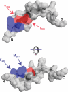
Spatial modeling of phenotypically distinctive YopD variants. Within the structure of the YopD C-terminal amphipathic domain (Protein Data Bank accession code: 1KDL) (49), the molecular surface positioning of the side groups of Val284 and Ile288 are highlighted in red. When substituted for a residue of opposing properties, these variants are essentially indistinguishable from the full-length yopD null mutant (9, 11, 27), being defective for both yop regulatory control and Yop effector translocation. Highlighted in blue are the side groups of Phe280 and Met281. When these are substituted for a residue of opposing characteristics, mutant YopD is unable to maintain yop regulatory control, although effector translocation remains essentially unaffected. The images display the molecular surface generated by SwissPdb Viewer (76). Images are related by a rotation out of the page about the horizontal axis by ∼90° to reveal two sides of the structure.
FIGURE 8.
FIGURE 8.
Binding of YopD variants to a GST-LcrV hybrid. In the Y. pseudotuberculosis ΔlcrVHyopBD background (YPIII/pIB191), gst alone (pGEX-5X-3) or translationally fused with lcrV (pMF777) was co-expressed under an IPTG-inducible promoter together with various yopD alleles (pMF781 and pTC006–pTC011) encoding for mutants harboring defined amino acid substitutions within the YopD amphipathic α-helix. Western blotting and the polyclonal α-YopD and monoclonal α-GST antibodies were used to detect the presence of YopD (∼33.3 kDa), GST (∼26 kDa), and GST-LcrV fusions (∼63 kDa) in the different fractions generated throughout the pulldown assay. Lanes: total bacterial cell lysate (C); pelleted insoluble fraction (I); soluble supernatant fraction (S); unbound material removed after collection of glutathione-Sepharose beads (U); material recovered from beads following first (W1), second (W2), third (W3), and fourth wash (W4); final bound material eluted from beads (B).
FIGURE 9.
FIGURE 9.
In vivo cross-linking of YopD. Various YopD derivatives were overexpressed in E. coli, and whole cell suspensions were cross-linked with the membrane-permeable cross-linker EGS (A and C) or DSP (B). Unless indicated otherwise (−), boiled total cell lysate was usually prepared in loading buffer with the addition of 5% (v/v) β-mercaptoethanol (β-ME) before being separated by SDS-PAGE and immunoblotted with polyclonal rabbit anti-YopD antiserum. Vector, E. coli containing the empty expression vector pET22b(+); and pYopD+, E. coli containing pET22b(+) carrying the wild type yopD allele (pMF839). Other YopD variants with substitutions of key residues in the C-terminal amphipathic α-helix (individual mutants indicated in subscript) were utilized in panel C. The numbers above each lane designate final concentrations (in mm) of each cross-linker. The positions of the protein standards with their approximate molecular mass (kDa) are marked at left. Asterisks at right mark protein bands appearing only in E. coli-expressing YopD and represent higher order YopD oligomers. Internal cross-linking of monomeric YopD most likely accounts for its retarded migration in the presence of increasing amounts of either cross-linker (¤).

Similar articles

Cited by

References

    1. Coburn B., Sekirov I., Finlay B. B. (2007) Clin. Microbiol. Rev. 20, 535–549 - PMC - PubMed
    1. Shao F. (2008) Curr. Opin. Microbiol. 11, 21–29 - PubMed
    1. Coombes B. K., Finlay B. B. (2005) Trends. Microbiol. 13, 92–95 - PubMed
    1. Blocker A., Holden D., Cornelis G. (2000) Cell. Microbiol. 2, 387–390 - PubMed
    1. Büttner D., Bonas U. (2002) Trends Microbiol. 10, 186–192 - PubMed

Publication types

MeSH terms

Substances