. 2010 Oct;38(19):6375-88.
doi: 10.1093/nar/gkq492. Epub 2010 Jun 4.

A role for YY1 in repression of dominant negative LEF-1 expression in colon cancer

Affiliations

A role for YY1 in repression of dominant negative LEF-1 expression in colon cancer

Noriko N Yokoyama et al. Nucleic Acids Res. 2010 Oct.

Abstract

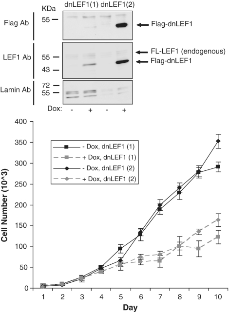
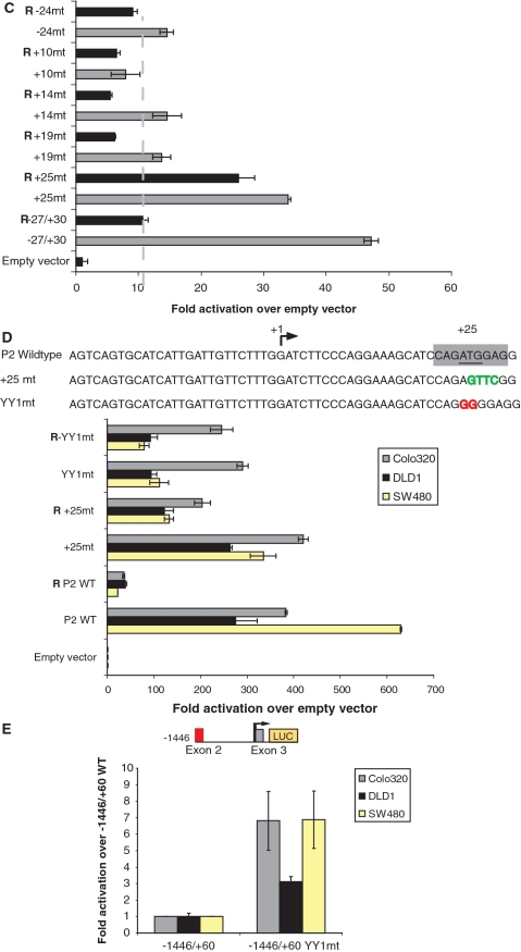
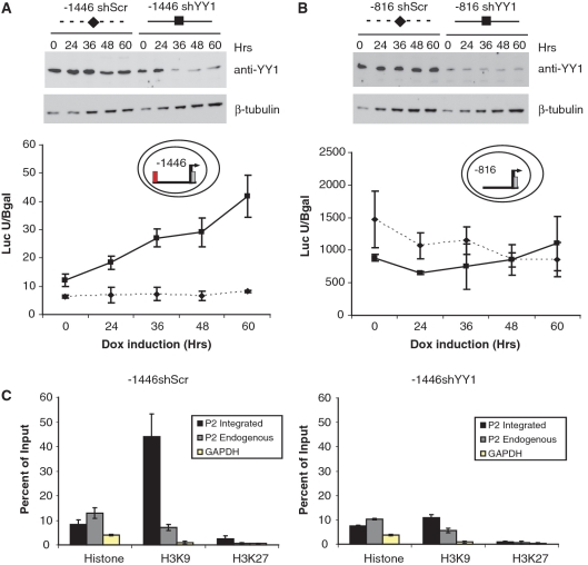
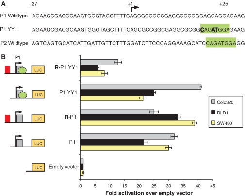
Lymphoid enhancer factor 1 (LEF-1) mediates Wnt signaling via recruitment of β-catenin to target genes. The LEF1 gene is aberrantly transcribed in colon cancers because promoter 1 (P1) is a Wnt target gene and is activated by TCF-β-catenin complexes. A second promoter in intron 2 (P2) produces dominant negative LEF-1 isoforms (dnLEF-1), but P2 is silent because it is repressed by an upstream distal repressor element. In this study we identify Yin Yang 1 (YY1) transcription factor as the P2-specific factor necessary for repression. Site-directed mutagenesis and EMSA were used to identify a YY1-binding site at +25 in P2, and chromatin immunoprecipitation assays detected YY1 binding to endogenous LEF1 P2. Mutation of this site relieves P2 repression in transient transfections, and knockdown of endogenous YY1 relieves repression of integrated P2 reporter constructs and decreases the H3K9me3 epigenetic marks. YY1 is responsible for repressor specificity because introduction of a single YY1-binding site into the P1 promoter makes it sensitive to the distal repressor. We also show that induced expression of dnLEF-1 in colon cancer cells slows their rate of proliferation. We propose that YY1 plays an important role in preventing dnLEF-1 expression and growth inhibition in colon cancer.


Figures

Figure 1.
Figure 1.
Two promoters in the LEF1 locus. The LEF1 locus contains two RNA polymerase II promoters. One promoter (P1) produces an mRNA that encodes full-length LEF-1 protein. A second promoter in intron 2 (P2) produces an mRNA that encodes the shorter, dominant negative LEF-1 isoform. Both promoters contain bona fide WREs (blue WREs) that mediate activation by TCF–β-catenin complexes. P1 and P2 expression patterns are aberrant in colon cancer cells. P1 is aberrantly activated by TCF–β-catenin, whereas P2 is silenced by a distal repressor located between –1446 and –1281 nt upstream relative to the P2 transcription start site (red circle). The repressor targets a unique feature of basal P2 as P1 is not silenced by the repressor.
Figure 2.
Figure 2.
Overexpression of dnLEF-1 slows down DLD-1 colon cancer cell growth. The effect of doxycycline-induced expression of dnLEF-1 on DLD-1 colon cancer cell growth was monitored over a 10-day period in two different clonal stable cells lines [dnLEF1(1) and dnLEF1(2)]. Quantitation of cell number with or without doxycycline (0.01 µg/ml) was performed using a sulforhodamine B cell proliferation assay (colorimetric-based growth curve). Differences in the rate of cell growth appeared after 4 days after which the dnLEF-1 expressing cells grew at a significantly reduced rate. A representative graph is shown from two independent trials. Error bars depict SDs of the results obtained with eight replicates from one trial. The western blots above the cell growth assay show the induced levels of expression of flag-tagged dnLEF-1 protein after 24 h of doxycycline treatment. Endogenous LEF-1 is also detected with LEF1 antibody (FL-LEF-1). Lamin antisera is used for a loading control.
Figure 3.
Figure 3.
An element in core promoter P2 is necessary for repression. (A) The 165-nt distal repressor (red box) was fused to P2 at different upstream positions (from –177 to –27 relative to the start site of transcription). Promoter activities were analyzed by luciferase reporter assays in Colo320 colon cancer cells. All promoter fragments were sensitive to repression, therefore, the repressor specifically targets core P2 sequences between –27 and +30. Data are derived from duplicate samples, and the results shown represent one of four replicate experiments. Fold activation was calculated as a ratio of luciferase levels from each reporter construct relative to the promoterless reporter vector. Error bars represent the spread between duplicate samples. (B) Multiple mutations of core P2 were generated by site-directed mutagenesis. P2 sequences from –27 to +30 are shown. Each mutation was introduced into pairs of luciferase reporter plasmids that differed only by the presence of the distal repressor element at –27 (pGL2E R–27/+30 and pGL2E –27/+30). Black bolded nucleotides show the essential initiator sequence (–17) and the transcription start site (+1). Green bolded nucleotides show the five different mutated sequences. (C) Luciferase assays of the mutated P2 reporter plasmids are shown. P2 reporter plasmids were transiently transfected into Colo320 cells. Mutations of core promoter sequences reduced P2 activity in all cases (gray bars), whereas only the mutation at +25 relieved repression by the distal repressor (black bars). Luciferase activity is reported as activation over that for a promoterless reporter vector. Data are derived from duplicate samples, and the results shown represent one of three replicate experiments. Error bars represent the spread between duplicate samples. (D) A mutation to destroy the YY1-binding site at +24 and +25 (AT to GG, in red) was introduced by site-directed mutagenesis. The mutation was introduced in luciferase reporter plasmid pGL2E R−27/+30 and pGL2E −27/+30 (P2 WT and R P2 WT respectively). Wild-type P2 and mutated P2 reporter plasmids were transiently transfected into three different colon cancer cell lines (Colo320, DLD-1 and SW480). Data shown are from a representative experiment out of three trials; error bars represent the spread of duplicate samples. The mutation at the YY1-binding site resulted in partial relief of repression in all three cell lines. (E) A YY1-binding site mutation was also introduced in the more physiologically relevant reporter plasmid pGL2B −1446/+60. Both wild-type −1446/+60 P2 and −1446/+60 YY1mt reporter plasmids were transiently transfected into three different colon cancer cell lines (Colo320, DLD-1, and SW480). Data are derived from three replicate experiments and error bars represent SDs of the data. For all three cell lines, introduction of the YY1-binding site mutation showed relief of P2 repression.
Figure 3.
Figure 3.
An element in core promoter P2 is necessary for repression. (A) The 165-nt distal repressor (red box) was fused to P2 at different upstream positions (from –177 to –27 relative to the start site of transcription). Promoter activities were analyzed by luciferase reporter assays in Colo320 colon cancer cells. All promoter fragments were sensitive to repression, therefore, the repressor specifically targets core P2 sequences between –27 and +30. Data are derived from duplicate samples, and the results shown represent one of four replicate experiments. Fold activation was calculated as a ratio of luciferase levels from each reporter construct relative to the promoterless reporter vector. Error bars represent the spread between duplicate samples. (B) Multiple mutations of core P2 were generated by site-directed mutagenesis. P2 sequences from –27 to +30 are shown. Each mutation was introduced into pairs of luciferase reporter plasmids that differed only by the presence of the distal repressor element at –27 (pGL2E R–27/+30 and pGL2E –27/+30). Black bolded nucleotides show the essential initiator sequence (–17) and the transcription start site (+1). Green bolded nucleotides show the five different mutated sequences. (C) Luciferase assays of the mutated P2 reporter plasmids are shown. P2 reporter plasmids were transiently transfected into Colo320 cells. Mutations of core promoter sequences reduced P2 activity in all cases (gray bars), whereas only the mutation at +25 relieved repression by the distal repressor (black bars). Luciferase activity is reported as activation over that for a promoterless reporter vector. Data are derived from duplicate samples, and the results shown represent one of three replicate experiments. Error bars represent the spread between duplicate samples. (D) A mutation to destroy the YY1-binding site at +24 and +25 (AT to GG, in red) was introduced by site-directed mutagenesis. The mutation was introduced in luciferase reporter plasmid pGL2E R−27/+30 and pGL2E −27/+30 (P2 WT and R P2 WT respectively). Wild-type P2 and mutated P2 reporter plasmids were transiently transfected into three different colon cancer cell lines (Colo320, DLD-1 and SW480). Data shown are from a representative experiment out of three trials; error bars represent the spread of duplicate samples. The mutation at the YY1-binding site resulted in partial relief of repression in all three cell lines. (E) A YY1-binding site mutation was also introduced in the more physiologically relevant reporter plasmid pGL2B −1446/+60. Both wild-type −1446/+60 P2 and −1446/+60 YY1mt reporter plasmids were transiently transfected into three different colon cancer cell lines (Colo320, DLD-1, and SW480). Data are derived from three replicate experiments and error bars represent SDs of the data. For all three cell lines, introduction of the YY1-binding site mutation showed relief of P2 repression.
Figure 4.
Figure 4.
YY1 binds to the +25 region of P2 in vitro and in vivo. (A) EMSA was performed with a 32P-labeled P2 probe containing the putative YY1 wild-type sequence (-CAGATGGAG-) or a mutated sequence (-CAGCGTGAG-) and purified recombinant human His-tagged YY1 protein. Purified His-tagged YY1 is shown in both Coomassie stained SDS–PAGE gel and YY1 probed western blot. Protein was added in increasing amounts (0.05, 0.1, 0.15 and 0.3 μg) (left EMSA). To confirm binding specificity, 1, 2 or 4 µg YY1 monoclonal antibody was included in the EMSA assay with 0.15 μg purified YY1 protein and 32P-labeled P2 probe (right EMSA). YY1 antibody blocked formation of the larger complex, but not the faster migrating complex. Arrows point to the YY1 bound probe. (B) ChIP assay was performed with Colo320 cells lysates. Chromatin from formaldehyde-crosslinked Colo320 cells was immunoprecipitated with monoclonal YY1 and pan-specific Histone antibodies. ChIP immunoprecipitates were analyzed by PCR primers that detect the core P2 region (−188 to +144), intron3 of the LEF1 gene, and an unrelated region of the genome (ZNF609). Data are derived from two to three independent experiments and error bars represent standard deviations. The difference between YY1 and IgG occupancy was significant for the core P2 region (P < 0.05) but insignificant for intron3 or ZNF609 (Student’s t test). Representative gels for each primer set are shown.
Figure 5.
Figure 5.
YY1 knockdown relieves P2 repression. Inducible shRNA YY1 (shYY1) and shRNA Scrambled (shScr) stable cell lines were derived from (A) Colo320 −1446/+60 and (B) −816/+60 luciferase reporter lines (‘Materials and Methods' section). Knockdown was induced with 1 µg/ml of doxycycline and cells were harvested 0, 24, 36, 48 and 60 h after treatment and processed for western blot analysis and luciferase assays. Doxycycline induction of YY1-specific shRNA leads to knockdown of YY1 protein (western insets; –1446 shYY1 and –816 shYY1), whereas induction of a scrambled, negative control shRNA has no effect (−1446/+60 shScr and −816/+60 shScr). Anti β-tubulin antibody was used to control for loading in the western blot analysis. All four cell lines carry LEF1 P2 luciferase and CMV-β-galactosidase reporter plasmids integrated in the cellular genome, therefore, we assayed extracts from the doxycycline-treated cells for both luciferase and β-galactosidase activities. β-galactosidase activity was used to normalize luciferase light units. The data are represented as ratios of luciferase light units to β-galactosidase units, and the error bars reflect standard deviation of the data from three independent assays. (C) ChIP assay was performed with −1446/+60 reporter integrated Colo320 stable cells with induction of either shScr or shYY1 plasmid to reduce YY1 protein levels. Chromatin from formaldehye-crosslinked cells (either shScr or shYY1) was immunoprecipitated with pan-specific Histone, polyclonal H3K9me3, monoclonal H3K27me3 and IgG antibodies. ChIP immunoprecipitates were analyzed by qPCR primers that detect integrated P2 (−108 to +60), endogenous P2 (−108 to +82), and primer set that detects the promoter of the GAPDH gene. Data are derived from two independent ChIP experiments and qPCR was performed in triplicate.
Figure 6.
Figure 6.
Introduction of a YY1-binding site confers repressor sensitivity to Promoter 1. (A) Wild-type P1 sequences (−27/+30) and a mutant P1 with an introduced YY1-binding site (boxed in green) are shown. For comparison, wild-type sequence of core P2 with its native YY1-binding site is shown. Site-directed mutagenesis was used to introduce the YY1-binding site into core P1 between +19 and +22 of two luciferase reporter plasmids that differed only by the presence and absence of the distal repressor element (P1 YY1 and R–P1 YY1). (B) Transient transfections of the indicated reporter plasmids were performed in Colo320, DLD-1 and SW480 colon cancer cell lines. The distal repressor element is indicated by a red box, the introduced YY1-binding site by a green oval and the core P1 promoter by a grey box. Luciferase activity is reported as fold activation over that for a promoterless reporter vector. Data are derived from duplicate samples, and the results shown are a representative of at least three experiments. Error bars represent the spread of duplicate samples.

Similar articles

Cited by

References

    1. Korinek V, Barker N, Morin PJ, van Wichen D, de Weger R, Kinzler KW, Vogelstein B, Clevers H. Constitutive transcriptional activation by a beta-catenin-Tcf complex in APC-/- colon carcinoma. Science. 1997;275:1784–1787. - PubMed
    1. Morin PJ, Sparks AB, Korinek V, Barker N, Clevers H, Vogelstein B, Kinzler KW. Activation of beta-catenin-Tcf signaling in colon cancer by mutations in beta-catenin or APC. Science. 1997;275:1787–1790. - PubMed
    1. Rubinfeld B, Robbins P, El-Gamil M, Albert I, Porfiri E, Polakis P. Stabilization of beta-catenin by genetic defects in melanoma cell lines. Science. 1997;275:1790–1792. - PubMed
    1. Li TW, Ting JH, Yokoyama NN, Bernstein A, van de Wetering M, Waterman ML. Wnt activation and alternative promoter repression of LEF1 in colon cancer. Mol. Cell Biol. 2006;26:5284–5299. - PMC - PubMed
    1. Behrens J, von Kries JP, Kuhl M, Bruhn L, Wedlich D, Grosschedl R, Birchmeier W. Functional interaction of beta-catenin with the transcription factor LEF-1. Nature. 1996;382:638–642. - PubMed

Publication types

MeSH terms

Substances