. 2010 Jul 1;185(1):190-202.
doi: 10.4049/jimmunol.0903505. Epub 2010 Jun 4.

Germinal center T follicular helper cell IL-4 production is dependent on signaling lymphocytic activation molecule receptor (CD150)

Affiliations

Germinal center T follicular helper cell IL-4 production is dependent on signaling lymphocytic activation molecule receptor (CD150)

Isharat Yusuf et al. J Immunol. .

Abstract

CD4 T cell help is critical for the generation and maintenance of germinal centers (GCs), and T follicular helper (T(FH)) cells are the CD4 T cell subset required for this process. Signaling lymphocytic activation molecule (SLAM)-associated protein (SAP [SH2D1A]) expression in CD4 T cells is essential for GC development. However, SAP-deficient mice have only a moderate defect in T(FH) differentiation, as defined by common T(FH) surface markers. CXCR5(+) T(FH) cells are found within the GC, as well as along the boundary regions of T/B cell zones. In this study, we show that GC-associated T follicular helper (GC T(FH)) cells can be identified by their coexpression of CXCR5 and the GL7 epitope, allowing for phenotypic and functional analysis of T(FH) and GC T(FH) populations. GC T(FH) cells are a functionally discrete subset of further polarized T(FH) cells, with enhanced B cell help capacity and a specialized ability to produce IL-4 in a T(H)2-independent manner. Strikingly, SAP-deficient mice have an absence of the GC T(FH) cell subset and SAP(-) T(FH) cells are defective in IL-4 and IL-21 production. We further demonstrate that SLAM (Slamf1, CD150), a surface receptor that uses SAP signaling, is specifically required for IL-4 production by GC T(FH) cells. GC T(FH) cells require IL-4 and -21 production for optimal help to B cells. These data illustrate complexities of SAP-dependent SLAM family receptor signaling, revealing a prominent role for SLAM receptor ligation in IL-4 production by GC CD4 T cells but not in T(FH) cell and GC T(FH) cell differentiation.


Conflict of interest statement

Disclosures

The authors have no financial conflicts of interest.

Figures

FIGURE 1
FIGURE 1
GC T cells (GC TFH) are distinct from TFH cells in their ability to produce IL-4. A and B, CD45.1+ SMtg CD4 T cells were adoptively transferred into C57BL/6 (B6)recipient mice subsequently infected with LCMV. A, SMtg CD4 T cells in spleen, day 8 postinfection. B, ICOS, PD-1, BTLA, and CXCR5 expression on SMtg cells; TFH cells are boxed. C, GL7 Ag and CXCR5 expression on nontransgenic CD44hiCD4 T cells, day 8 after LCMV infection. Non-TFH cells (CXCR5 −/lo), GL7TFH cells (CXCR5hiGL7), and GC TFH cells (CXCR5hiGL7+) are boxed. D, PD-1 expression on GC TFH, GL7TFH, non-TFH, and naive CD4 cells. Data are representative of at least three independent experiments. E, Immunofluorescence histology of spleen, 8 d after CD45.1+ OTII CD4 T cell transfer and NP-OVA immunization. GL7 (green), CD45.1 (red), and B220 (blue). CD45.1+ CD4 T cells inside the GC are marked by white boxes, and representative CD45.1+ CD4 T cells outside the GC are marked by yellow boxes. Enlarged views of the boxed CD4 T cells are shown to the right. Original magnification × 200. F, Quantitation of GL7+ and GL7CD45.1+ CD4 T cells outside GCs. GP, mRNA expression by polyclonal LCMV-specific CD44hi CD4 T cells, day 8 postinfection. Non-TFH (CXCR5lo), TFH (CXCR5hiGL7), and GC TFH (CXCR5hiGL7+) cells. Naive cells (CD4+CD44lo). Expression levels were quantitated in reference to β-actin mRNA and then normalized to naive CD4 T cells, except CXCR5, which was quantified by gene-expression microarray. G, Cmah. H, Bcl6. I, PD-1. J, ICOS. K, BTLA. L, CD200. M, IL-21. N, CXCR5. O, Prdm1/Blimp-1. P, IL-4. n = 2/group, with 10 spleens pooled per sample. **p < 0.01; ***p < 0.001.
FIGURE 2
FIGURE 2
LCMV-specific TFH cells and GC TFH cells exhibit TH1, but not TH2, attributes. A and B, Day 8 after LCMV infection of B6 mice that were recipients of SMtg CD4 T cells. IFN-γ expression by CXCR5 non-TFH cells and CXCR5+ TFH SMtg CD4 T cells after a 4-h in vitro restimulation with PMA/ionomycin. A, Gated SMtg CD4 T cells are shown. Boxes show non-TFH and TFH cell gates. B, IFN-γ expression by cells gated in A (n = 5/group). Data are representative of three independent experiments. T-bet (C) and IFN-γ (D) mRNA expression by polyclonal LCMV-specific CD44hi CD4+ T cells, 8 d after LCMV infection, directly ex vivo. Normalized as in Fig. 1. ***p < 0.001. E, Serum Ab levels (μg/ml) of IgG2c and IgG1 in LCMV-infected B6 mice. GATA3 (F) and c-Maf (G) mRNA expression, quantitated as above.
FIGURE 3
FIGURE 3
IL-4 production by GC TFH cells but not TFH or non-TFH cells. IL-4 production was tracked in 4 get IL-4/GFP reporter mice after LCMV infection. A, IL-4 production by CXCR5+ CD4 T cells. Gated CD44hi CD4 T cells are shown. B, CXCR5+ CD4 T cells were then gated on the basis of GL7 expression (GL7+ GC TFH, GL7 TFH, as per Fig. 1C), and GFP+ IL-4 producing cells were quantified. C, Antiviral CD44hi CD4 T cells were gated on the basis of CXCR5 expression, and GFP+ IL-4–producing cells in each population were quantified. **p < 0.01; ***p < 0.001.
FIGURE 4
FIGURE 4
SAP mice have a reduced TFH population after LCMV infection. A, SAP/SH2D1A mRNA expression in polyclonal LCMV-specific CD44hi CD4+ non-TFH cells (CXCR5−/lo), TFH cells (CXCR5hi GL7), and GC TFH cells (CXCR5hi GL7+), day 8 after LCMV infection. Expression levels normalized as in Fig. 1. B, SAP protein expression in SAP naive (CD44lo), WT naive (CD44lo), GC TFH (CD44hiCXCR5hiGL7+), TFH (CD44hiCXCR5hiGL7), and non-TFH (CD44hi CXCR5−/lo) CD4 T cells by flow cytometry. C, Quantitation of SAP protein mean fluorescence intensity (MFI) from B. Data are representative of two independent experiments (n = 6). DG, WT or SAP SMtg CD4 T cells were adoptively transferred into B6 recipient mice subsequently infected with LCMV. Flow cytometric analysis of ICOS (D), PD-1 (E), and BTLA (F), gated on SMtg CD4 T cells at day 8 postinfection. G, SMtg TFH cell quantitation. Data are representative of three experiments. H, TFH differentiation of polyclonal LCMV-specific (CD44hi) CD4+ T cells in WT and SAP mice, 8 d postinfection. Naive (CD44lo) CD4 T cells are also shown. I, GC B cell frequency in WT and SAP- mice, 8 d after LCMV infection. Data are representative of >10 independent experiments. J, WT or SAP OTII CD4 T cells were adoptively transferred into B6 recipient mice subsequently immunized with NP-OVA. CXCR5 expression on naive CD44lo B6 CD4 T cells, WT OTII cells, and SAP OTII CD4+ T cells. K, Quantitation of TFH data in J. Data are representative of four independent experiments. **p < 0.01; ***p < 0.001.
FIGURE 5
FIGURE 5
SAP is required for GC TFH cell differentiation. A, B, GC TFH cell differentiation in WT or SAP mice after LCMV infection. A, Flow cytometry of GC TFH cell frequencies (CXCR5hi GL7+). Gated CD44hi CD4 T cells are shown. B, GC TFH cell quantitation from data in A (n = 5/group). Data are representative of three independent experiments. CE, Congenically marked WT or SAP OTII CD4 T cells were transferred into B6 mice subsequently immunized with NP-OVA. Eight days later, OTII CD4 T cells were analyzed for GC TFH cell differentiation. C, Total CD4 T cells are shown. Box shows OTII cell gate. D, GC TFH cell differentiation by WT and SAP OTII cells. CD44hiCD62LloCD4+ T cells are shown. GC TFH cells are boxed. E, GC TFH cell percentage of OTII CD44hiCD62Llo CD4 T cells (n = 4/group). Data are representative of three independent experiments. F, PD-1 expression by WT and SAP TFH cells. ***p < 0.001.
FIGURE 6
FIGURE 6
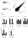
SAP-deficient TFH cells do not express IL-4. WT or SAP SMtg CD4 T cells were adoptively transferred into B6 recipient mice subsequently infected with LCMV. A, WT and SAP TFH SMtg CD4 T cells were sorted by FACS on the basis of CXCR5 expression at day 8 postinfection. TFH cell phenotype was confirmed by staining for CXCR5hiSLAMlo expression on reserved aliquots of cells. Isolated mRNA from sorted cells was used for gene-expression microarray. B, Scatter plot of WT TFH cell versus SAP TFH cell microarray gene-expression data, log 2 scale. Black lines indicate 3-fold expression changes. R2 = 0.9905; 45,101 gene probes shown. Average signal of biological replicates is plotted. Data are shown from one of two independent experiments (n = 2/group). CH, Gene expression in naive (CD44lo, from uninfected mice), WT non-TFH (CXCR5−/lo), SAP non-TFH (CXCR5−/lo), WT TFH (CXCR5hi), and SAP TFH (CXCR5hi) SMtg CD4 T cells, by Affymetrix microarray (n = 2/group). Each sample consisted of cells from 10 pooled spleens. mRNA expression of each gene was normalized to naive CD4 T cell controls. C, Bcl6. D, Prdm1/Blimp-1. E, ICOS. F, CD200. G, IL-21. H, IL-4.
FIGURE 7
FIGURE 7
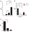
SLAM-deficient GC TFH cells exhibit impaired IL-4 production. A, Flow cytometric analysis of SLAM expression on naive CD4 T cells and in vitro polarized TH1 and TH2 CD4 T cells. B, Flow cytometric analysis of SLAM and CXCR5 expression on CD45.1+ SMtg CD4 T cells, day 8 after LCMV infection. C, Microarray analysis of SLAM mRNA in naive (CD44lo, from uninfected mice), non-TFH (CXCR5lo), and TFH (CXCR5hi) SMtg CD4 T cells. D and E, Flow cytometry on splenocytes, day 8 after LCMV infection of B6 mice. D, SLAM expression on naive (CD44lo), non-TFH (CD44hi CXCR5lo), TFH (CD44hiCXCR5loGL7), and GC TFH (CD44hiCXCR5+GL7+) endogenous CD4 T cells. E, SLAM mean fluorescence intensity (MFI), quantified from experiments in D. F, SLAM MFI on IgD+ (naive) and GC B cells, day 8 after LCMV infection of B6 mice. Data are representative of three to five independent experiments. G, GC TFH (CXCR5hiGL7+), TFH (CXCR5hiGL7), and non-TFH (CXCR5−/lo) CD4 T cell populations in Slamf1−/− (SLAM−/−) and Slamf1+/+ (SLAM+/+) mice, 8 d after LCMV infection. Gated CD44hiCD4+ T cells are shown. Quantitated TFH cell (H) and GC TFH cell (I) frequencies in SLAM+/+ and SLAM−/− mice, from experiment shown in G (n = 6/group). Data are representative of three independent experiments. J and K, GC TFH, TFH, and non-TFH CD4 T cells, as gated in G, were sorted from SLAM −/− and SLAM+/+ mice, 8 d after LCMV infection. Expression of IL-4 (J) and IL-21 (K) mRNA in SLAM+/+ and SLAM−/− GC TFH cells. Expression levels by qPCR were quantitated in reference to a control gene and then normalized to naive CD4 T cells. Data are representative of two experiments (n = 3/group). **p < 0.01; ***p < 0.001.
FIGURE 8
FIGURE 8
IL-4 is needed for optimal GC TFH B cell help. A, Non-TFH (CD44hiCXCR5loGL7), TFH (CD44hiCXCR5hiGL7), and GC TFH (CD44hiCXCR5+GL7+) CD4 T cells were sorted from spleens of LCMV-infected mice and cultured with purified B cells. IgG was quantitated in day 7 culture supernatants by ELISA. Data are representative of four experiments (n = 3/group). B, TFH (CD44hiCXCR5hiGL7) and GC TFH (CD44hiCXCR5+GL7+) CD4 T cells were sorted from spleens of LCMV-infected mice and cultured with purified B cells in the presence or absence of αIL4 or αIL21 mAb. IgG was quantitated in day 7 culture supernatants by ELISA. Data are representative of two experiments (n = 3/group). C, GC TFH (CD44hiCXCR5+GL7+) CD4 T cells were sorted from SLAM−/− and SLAM+/+ LCMV-infected mice and then cocultured with purified B cells. IgG was quantitated in day 7 culture supernatants by ELISA. Data are representative of two experiments (n = 3/group). *p < 0.05; **p < 0.01; ***p < 0.001.
FIGURE 9
FIGURE 9
Two-stage model of TFH cell differentiation, showing the regulation and impact of SLAM and SAP on TFH and GC TFH cells.

Similar articles

Cited by

References

    1. Schoenberger SP, Crotty S. Immunologic memory. In: Paul WE, editor. Fundamental Immunology. 6. Lippincott Williams & Wilkins; Philadelphia: 2008. pp. 862–898.
    1. McHeyzer-Williams M. B Lymphocyte biology. In: Paul WE, editor. Fundamental Immunology. 6. Lippincott Williams & Wilkins; Philadelphia: 2008. pp. 289–311.
    1. Amanna IJ, Slifka MK, Crotty S. Immunity and immunological memory following smallpox vaccination. Immunol Rev. 2006;211:320–337. - PubMed
    1. Radbruch A, Muehlinghaus G, Luger EO, Inamine A, Smith KG, Dörner T, Hiepe F. Competence and competition: the challenge of becoming a long-lived plasma cell. Nat Rev Immunol. 2006;6:741–750. - PubMed
    1. Tangye SG, Tarlinton DM. Memory B cells: effectors of long-lived immune responses. Eur J Immunol. 2009;39:2065–2075. - PubMed

Publication types

MeSH terms

Substances

Associated data