. 2010 Jul 1;185(1):183-9.
doi: 10.4049/jimmunol.0903846. Epub 2010 Jun 4.

Human T-lymphotropic virus type 1-induced CC chemokine ligand 22 maintains a high frequency of functional FoxP3+ regulatory T cells

Affiliations

Human T-lymphotropic virus type 1-induced CC chemokine ligand 22 maintains a high frequency of functional FoxP3+ regulatory T cells

Frederic Toulza et al. J Immunol. .

Abstract

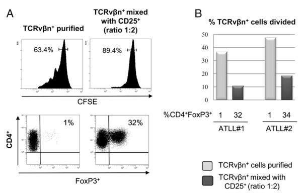
We recently reported that human T-lymphotropic virus type 1 (HTLV-1) infection is accompanied by a high frequency of CD4(+)FoxP3(+) cells in the circulation. In asymptomatic carriers of HTLV-1 and in patients with HTLV-1-associated inflammatory and malignant diseases, a high FoxP3(+) cell frequency correlated with inefficient cytotoxic T cell-mediated killing of HTLV-1-infected cells. In adult T cell leukemia/lymphoma (ATLL), the FoxP3(+) population was distinct from the leukemic T cell clones. However, the cause of the increase in FoxP3(+) cell frequency in HTLV-1 infection was unknown. In this study, we report that the plasma concentration of the chemokine CCL22 is abnormally high in HTLV-1-infected subjects and that the concentration is strongly correlated with the frequency of FoxP3(+) cells, which express the CCL22 receptor CCR4. Further, we show that CCL22 is produced by cells that express the HTLV-1 transactivator protein Tax, and that the increased CCL22 enhances the migration and survival of FoxP3(+) cells in vitro. Finally, we show that FoxP3(+) cells inhibit the proliferation of ex vivo, autologous leukemic clones from patients with ATLL. We conclude that HTLV-1-induced CCL22 causes the high frequency of FoxP3(+) cells observed in HTLV-1 infection; these FoxP3(+) cells may both retard the progression of ATLL and HTLV-1-associated inflammatory diseases and contribute to the immune suppression seen in HTLV-1 infection, especially in ATLL.


Figures

FIGURE 1
FIGURE 1
CCL22 concentration in plasma and culture supernatant. Concentration of CCL22 in fresh plasma (A) or in PBMC supernatant after 18 h incubation in vitro (B). Median values are shown under each box plot. The value is expressed in picograms per milliliter for plasma samples (A) or in picograms per milliliter per 106 PBMCs (B). The p values were calculated using Student t test.
FIGURE 2
FIGURE 2
Tax expression and CCL22 expression. A, Expression of CCL22 and HTLV-1 Tax protein (left panel) and CCL22 and FoxP3 (right panel) in CD4+ cells from two independent representative HTLV-1–infected patients. B, Frequency in PBMCs of T cells of the following phenotypes: CCL22CD4+Tax+, CCL22+CD4+Tax+, and CD4+CCL22+. The last column shows the frequency of CCL22+ cells in the CD4+Tax+ population.
FIGURE 3
FIGURE 3
Modulation and induction of CCL22 expression. A, Concentration of CCL22 (picograms per milliliter per 106 PBMCs) in PBMC supernatant after 18 h incubation in vitro of whole PBMCs (left) and PBMCs depleted of CD8+ cells (right). Each line represents the rise in CCL22 concentration for each respective subject after CD8+ cell depletion. B, Concentration of CCL22 (picograms per milliliter per 106 PBMCs) detected in supernatant of Jurkat cells in the presence or absence of anti-CD3 coated beads and in the presence or absence of MT2 cells. C, Concentration of CCL22 (picograms per milliliter per 106 PBMCs) in supernatant of Jurkat cells transfected with different plasmids (5 μg/ml/106 cells). Supernatant was obtained 72 h after transfection. D, Correlation between the plasma concentration of CCL22 (picograms per milliliter) and the frequency of Tax expression in CD4+ T cells. The solid line represents the regression curve for patients with HAM/TSP and the dashed line represents the regression curve for ACs.
FIGURE 4
FIGURE 4
Proviral load is associated with CCL22 concentration in plasma. There was a significant positive correlation between the proviral load and the plasma concentration of CCL22 in patients with HAM/TSP (n = 10) and ACs (n = 8).
FIGURE 5
FIGURE 5
CD4+FoxP3+ frequency in nonleukemic HTLV-1–infected individuals correlates with CCL22 concentration in plasma and supernatant of PBMCs. Correlation between the frequency of circulating CD4+FoxP3+ cells and the concentration of CCL22 in plasma from uninfected controls, ACs, and patients with HAM/TSP (A) and in patients with acute and chronic ATLL (B). The p values were determined by a two-tailed Spearman test.
FIGURE 6
FIGURE 6
Functional effects of CCL22 on CD4+FoxP3+ cells of HTLV-1–infected patients. A, Rate of migration of CD4+FoxP3+ cells in a transwell assay. The results are expressed as the fold increase in the number of cells that migrated in response to different concentrations of CCL22, compared with medium alone, during 4 h incubation. The results represent the means of four independent experiments. B, Viability of each respective cell subset following 24h of incubation, according to the level of CCL22 chemokine added. The values represent the absolute number of viable cells, normalized to the control sample. The histogram represents the mean of four independent experiments.
FIGURE 7
FIGURE 7
FoxP3+ cells inhibit the proliferation of autologous ATLL cells in patients with chronic ATLL. A, upper panels, Expression of CFSE in purified TCRvβn+ clones from two patients with ATLL; values represent the percentage of undivided CD4+ cells. Lower panels, CD4 and FoxP3 expression in the CD4+ gated population. B, Percentage of CFSE-labeled leukemic (TCRvβn+) cells undergoing division during 4 d incubation in vitro in the presence of different percentages of autologous CD4+FoxP3+ cells.

References

    1. Osame M, Usuku K, Izumo S, Ijichi N, Amitani H, Igata A, Matsumoto M, Tara M. HTLV-I associated myelopathy, a new clinical entity. Lancet. 1986;1:1031–1032. - PubMed
    1. Nagai M, Osame M. Human T-cell lymphotropic virus type I and neurological diseases. J. Neurovirol. 2003;9:228–235. - PubMed
    1. Uchiyama T, Yodoi J, Sagawa K, Takatsuki K, Uchino H. Adult T-cell leukemia: clinical and hematologic features of 16 cases. Blood. 1977;50:481–492. - PubMed
    1. Miyoshi I, Kubonishi I, Yoshimoto S, Akagi T, Ohtsuki Y, Shiraishi Y, Nagata K, Hinuma Y. Type C virus particles in a cord T-cell line derived by co-cultivating normal human cord leukocytes and human leukaemic T cells. Nature. 1981;294:770–771. - PubMed
    1. Kalyanaraman VS, Sarngadharan MG, Nakao Y, Ito Y, Aoki T, Gallo RC. Natural antibodies to the structural core protein (p24) of the human T-cell leukemia (lymphoma) retrovirus found in sera of leukemia patients in Japan. Proc. Natl. Acad. Sci. USA. 1982;79:1653–1657. - PMC - PubMed

Publication types

MeSH terms