. 2010 Oct;38(19):6796-802.
doi: 10.1093/nar/gkq508. Epub 2010 Jun 4.

Reliable semi-synthesis of hydrolysis-resistant 3'-peptidyl-tRNA conjugates containing genuine tRNA modifications

Affiliations

Reliable semi-synthesis of hydrolysis-resistant 3'-peptidyl-tRNA conjugates containing genuine tRNA modifications

Dagmar Graber et al. Nucleic Acids Res. 2010 Oct.

Abstract

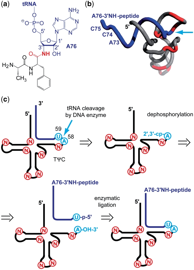
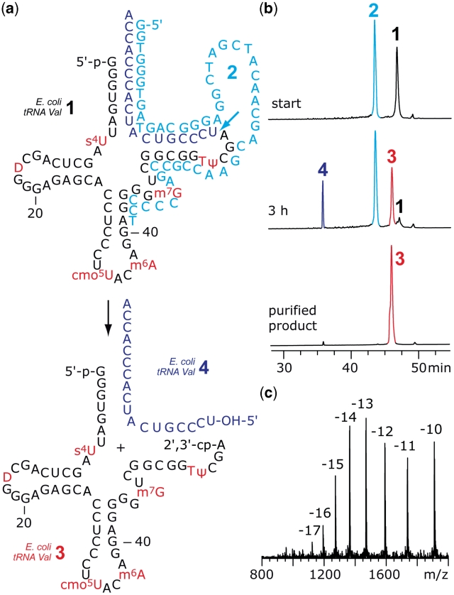
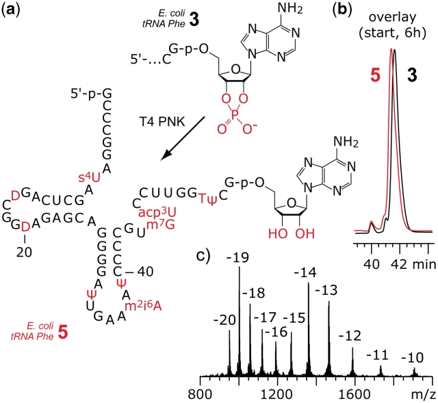
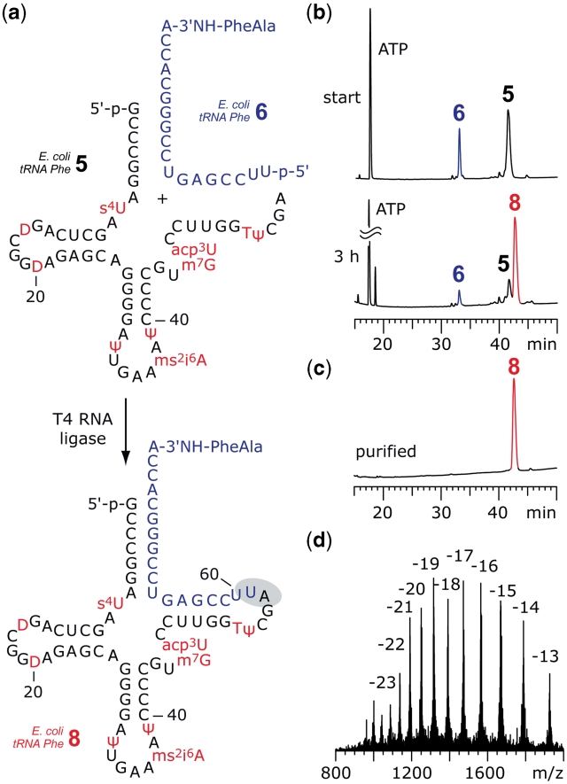
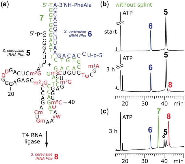
The 3'-peptidyl-tRNA conjugates that possess a hydrolysis-resistant ribose-3'-amide linkage instead of the natural ester linkage would represent valuable substrates for ribosomal studies. Up to date, access to these derivatives is severely limited. Here, we present a novel approach for the reliable synthesis of non-hydrolyzable 3'-peptidyl-tRNAs that contain all the respective genuine nucleoside modifications. In short, the approach is based on tRNAs from natural sources that are site-specifically cleaved within the TΨC loop by using DNA enzymes to obtain defined tRNA 5'-fragments carrying the modifications. After dephosphorylation of the 2',3'-cyclophosphate moieties from these fragments, they are ligated to the respective 3'-peptidylamino-tRNA termini that were prepared following the lines of a recently reported solid-phase synthesis. By this novel concept, non-hydrolyzable 3'-peptidyl-tRNA conjugates possessing all natural nucleoside modifications are accessible in highly efficient manner.


Figures

Figure 1.
Figure 1.
Hydrolysis-resistant 3′-peptidyl-tRNA conjugates. (a) Crucial structural feature of an exemplary target: The amide linkage (highlighted in red). (b) Cartoon presentation of a 3′-peptidyl-tRNA conjugate. Typical positions of natural chemical modifications are shown in red (PDB coordinates 1EVV: S. cerevisiae tRNAPhe). The 3′-terminal sequences (18 nt) of tRNAs are generally unmodified (blue); arrow (cyan) indicates the intended site of tRNA cleavage (by using DNA enzymes) and the site of ligation (to synthetic 3′-peptidylamino-RNA conjugates); (c) concept for the semi-synthesis of non-hydrolyzable 3′-peptidyl-tRNA conjugates (for explanation see main text). Modified nucleosides, N; cyclophosphate, cp; phosphate, p.
Figure 2.
Figure 2.
Example for the cleavage of a natural tRNA by DNA enzymes. (a) Secondary structure of E. coli tRNAVal 1 and 10–23 DNA enzyme 2. The desired tRNA 5′-fragment 3 contains all modified nucleosides and possesses a 2′,3′-cyclophosphate terminus. (b) Anion-exchange HPLC analysis of the cleavage reaction (88% yield according to peak area integration) and the purified fragment; reaction conditions: cDNAzyme = 80 µM; ctRNA = 40 µM; 50 mM Tris–HCl (pH 7.5), 10 mM MgCl2, 10 mM DTT, temperature cycles (25–90°C); anion-exchange HPLC: for conditions see ‘Materials and Methods’ section. (c) LC-ESI MS analysis of 3: m.w. (calcd) = 19113, m.w. (found) = 19112 ± 10. For structures and abbreviations of modified nucleosides, see Supplementary Data.
Figure 3.
Figure 3.
Example for enzymatic dephosphorylation of the 2′,3′-cyclophosphate at the tRNA 5′-fragment. (a) Structure of the 5′-fragment from E. coli tRNAPhe 2′,3′-cyclophosphate 3 and product 5 after exposure to T4 PNK. (b) The PNK reaction was monitored by anion-exchange HPLC analysis. The difference in retention time between 3 and 5 is marginal; reaction conditions: T4 PNK (0.5 U/µl; cRNA = 15 µM; 70 mM Tris–HCl (pH 7.6), 10 mM MgCl2, 5 mM DTT, 37°C. (c) LC-ESI MS analysis of 5: m.w. (calcd) = 19045, m.w. (found) = 19046 ± 10. Anion-exchange HPLC: for conditions see ‘Materials and Methods’ section. For structures and abbreviations of modified nucleosides see Supplementary Data.
Figure 4.
Figure 4.
Example for enzymatic ligation of fully modified tRNA 5′-fragments to synthetic 3′-peptidylamino-RNA conjugates. (a) Structures of the 5′-fragment from E. coli tRNAPhe 5 and the dipeptide-RNA conjugate 6 to form a preligation complex that allows T4 RNA ligation of the full-length tRNA-peptide conjugate 8. (b) The ligation reaction was monitored by anion-exchange HPLC analysis: 83% yield was achieved after 3 h; reaction conditions: T4 RNA ligase (0.5 U/µl; cRNA = 40 µM each strand; donor/acceptor = 1/1), 50 mM HEPES–NaOH (pH 8.0), 10 mM MgCl2, 10 mM DTT, 1 mM ATP, 0.1 mg/ml BSA, 37°C. (c) Purified 3′-peptidyl-tRNA; (d) LC-ESI MS analysis of 8: m.w. (calcd) = 25030, m.w. (found) = 25029 ± 10. Anion-exchange HPLC: for conditions see ‘Materials and Methods’ section. For structures and abbreviations of modified nucleosides see Supplementary Data.
Figure 5.
Figure 5.
Example for splint-assisted enzymatic ligation of fully modified tRNA 5′-fragments to synthetic 3′-peptidylamino-RNA conjugates. (a) Structures of the 5′-fragment from S. cerevisiae tRNAPhe 5 and the dipeptide-RNA conjugate 6 to form a preligation complex that allows T4 RNA ligation of the full-length tRNA-peptide conjugate 8. (b) Without splint 7 only marginal amounts of product 8 were formed; reaction conditions: T4 RNA ligase (0.5 U/µl; cRNA = 40 µM each strand; donor/acceptor = 1/1), 50 mM HEPES–NaOH (pH 8.0), 10 mM MgCl2, 10 mM DTT, 1 mM ATP, 0.1 mg/ml BSA, 37°C. (c) Ligation promoted by splint 7 resulted in 75% yield of 8. The reaction was monitored by anion-exchange HPLC (for conditions see ‘Materials and Methods’ section); an unidentified, unreactive impurity is marked by an asterisk; reaction conditions: T4 RNA ligase (0.25 U/µl; cRNA = 40 µM each strand; cDNA = 40 µM; donor/acceptor/splint = 1/1/1), buffer as in (b) and 0.5 mM ATP, 37°C. For structures and abbreviations of modified nucleosides see Supplementart Data.

Similar articles

Cited by

References

    1. Wilson DN, Nierhaus KH. The ribosome through the looking glass. Angew. Chem. Int. Ed. Engl. 2003;42:3464–3486. - PubMed
    1. Voorhees RM, Weixlbaumer A, Loakes D, Kelley AC, Ramakrishnan V. Insights into substrate stabilization from snapshots of the peptidyl transferase center of the intact 70S ribosome. Nat. Struct. Mol. Biol. 2009;16:528–533. - PMC - PubMed
    1. Lang K, Erlacher M, Wilson DN, Micura R, Polacek N. The role of 23S ribosomal RNA residue A2451 in peptide bond synthesis revealed by atomic mutagenesis. Chem. Biol. 2008;15:485–492. - PubMed
    1. Zhang W, Dunkle JA, Cate JH. Structures of the ribosome in intermediate states of ratcheting. Science. 2009;325:1014–1017. - PMC - PubMed
    1. Munro JB, Altman RB, O’Connor N, Blanchard SC. Identification of two distinct hybrid state intermediates on the ribosome. Mol. Cell. 2007;25:505–517. - PMC - PubMed

Publication types