

Monte Carlo investigations of the effect of beam divergence on thick, segmented crystalline scintillators for radiotherapy imaging
- PMID: 20526032
- PMCID: PMC2909124
- DOI: 10.1088/0031-9155/55/13/006
Monte Carlo investigations of the effect of beam divergence on thick, segmented crystalline scintillators for radiotherapy imaging
Abstract
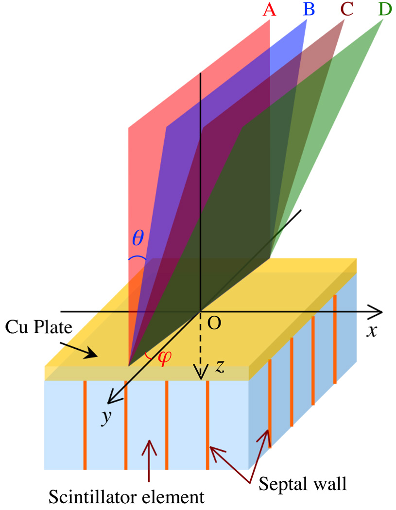
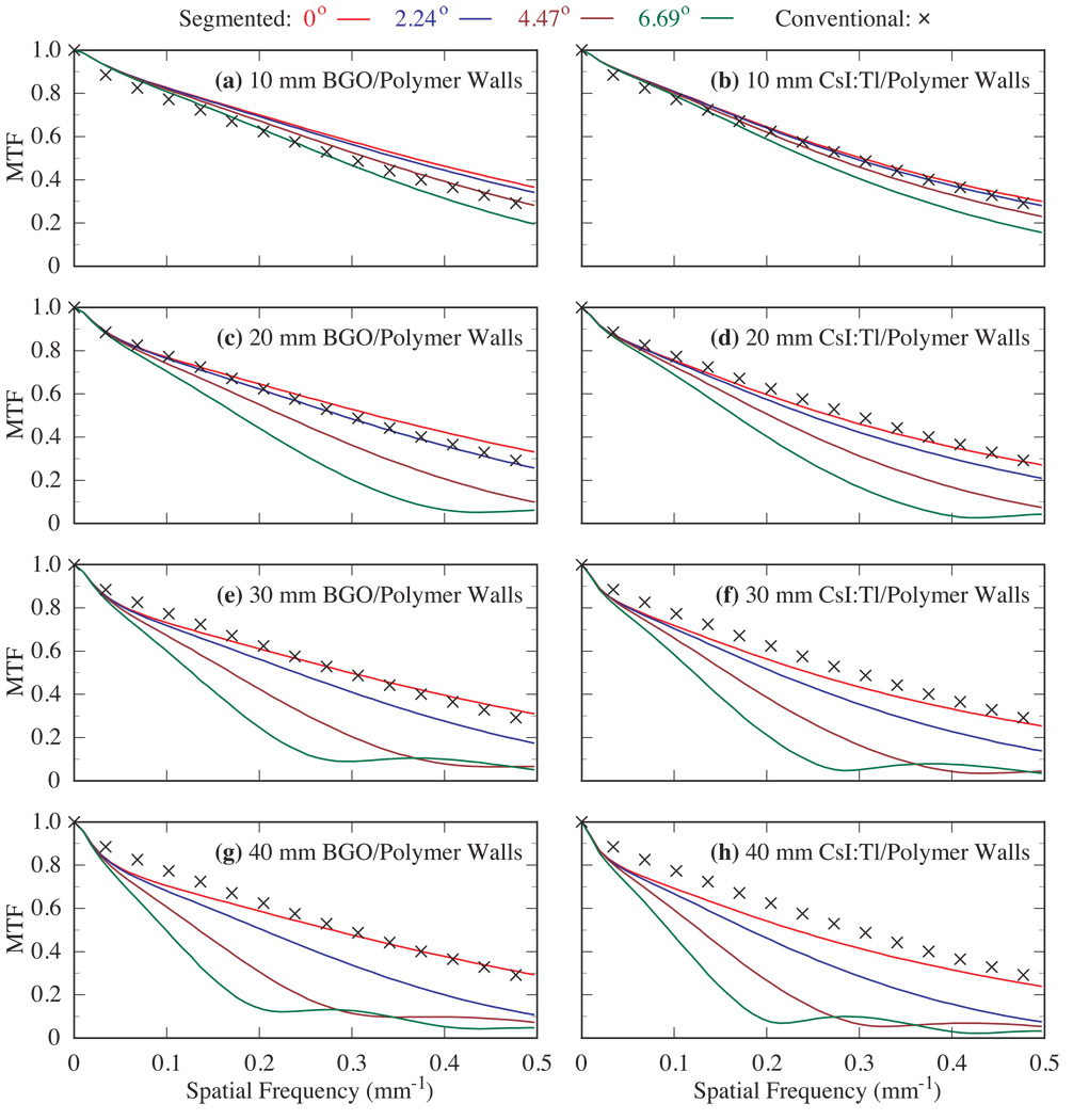
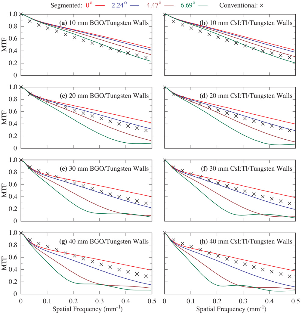
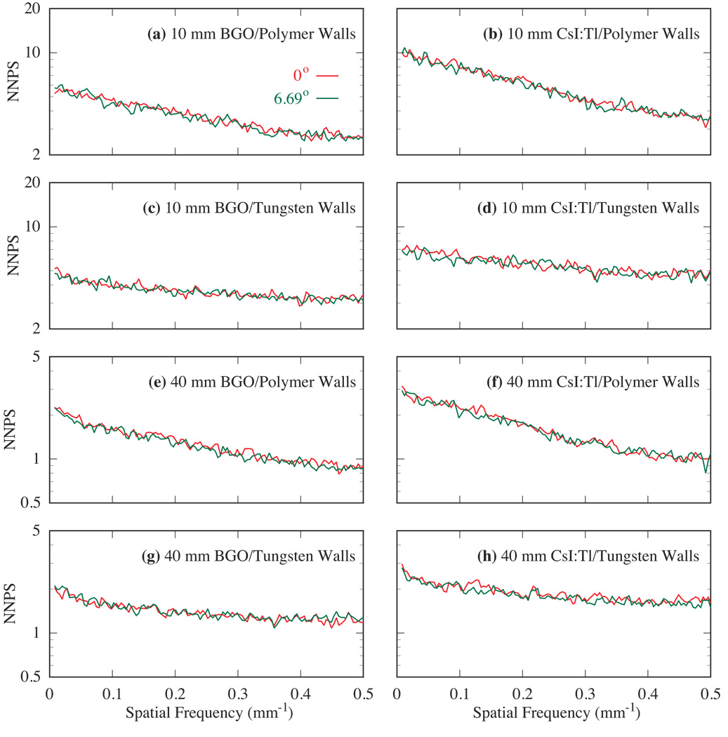
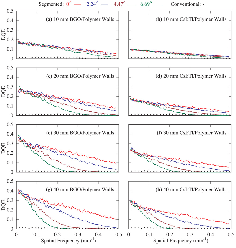
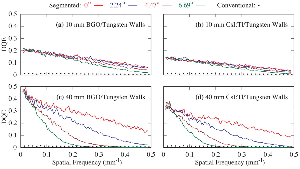
The use of thick, segmented scintillators in electronic portal imagers offers the potential for significant improvement in x-ray detection efficiency compared to conventional phosphor screens. Such improvement substantially increases the detective quantum efficiency (DQE), leading to the possibility of achieving soft-tissue visualization at clinically practical (i.e. low) doses using megavoltage (MV) cone-beam computed tomography. While these DQE increases are greatest at zero spatial frequency, they are diminished at higher frequencies as a result of degradation of spatial resolution due to lateral spreading of secondary radiation within the scintillator--an effect that is more pronounced for thicker scintillators. The extent of this spreading is even more accentuated for radiation impinging the scintillator at oblique angles of incidence due to beam divergence. In this paper, Monte Carlo simulations of radiation transport, performed to investigate and quantify the effects of beam divergence on the imaging performance of MV imagers based on two promising scintillators (BGO and CsI:Tl), are reported. In these studies, 10-40 mm thick scintillators, incorporating low-density polymer, or high-density tungsten septal walls, were examined for incident angles corresponding to that encountered at locations up to approximately 15 cm from the central beam axis (for an imager located 130 cm from a radiotherapy x-ray source). The simulations demonstrate progressively more severe spatial resolution degradation (quantified in terms of the effect on the modulation transfer function) as a function of increasing angle of incidence (as well as of the scintillator thickness). Since the noise power behavior was found to be largely independent of the incident angle, the dependence of the DQE on the incident angle is therefore primarily determined by the spatial resolution. The observed DQE degradation suggests that 10 mm thick scintillators are not strongly affected by beam divergence for detector areas up to approximately 30x30 cm2. For thicker scintillators, the area that is relatively unaffected is significantly reduced, requiring a focused scintillator geometry in order to preserve spatial resolution, and thus DQE.
Figures
Similar articles
-
Optimization of the design of thick, segmented scintillators for megavoltage cone-beam CT using a novel, hybrid modeling technique.Med Phys. 2014 Jun;41(6):061916. doi: 10.1118/1.4875724. Med Phys. 2014. PMID: 24877827 Free PMC article.
-
Countering beam divergence effects with focused segmented scintillators for high DQE megavoltage active matrix imagers.Phys Med Biol. 2012 Aug 21;57(16):5343-58. doi: 10.1088/0031-9155/57/16/5343. Epub 2012 Aug 1. Phys Med Biol. 2012. PMID: 22854009 Free PMC article.
-
Segmented crystalline scintillators: an initial investigation of high quantum efficiency detectors for megavoltage x-ray imaging.Med Phys. 2005 Oct;32(10):3067-83. doi: 10.1118/1.2008407. Med Phys. 2005. PMID: 16279059
-
High-DQE EPIDs based on thick, segmented BGO and CsI:Tl scintillators: performance evaluation at extremely low dose.Med Phys. 2009 Dec;36(12):5707-18. doi: 10.1118/1.3259721. Med Phys. 2009. PMID: 20095283 Free PMC article.
-
Segmented crystalline scintillators: empirical and theoretical investigation of a high quantum efficiency EPID based on an initial engineering prototype CsI(TI) detector.Med Phys. 2006 Apr;33(4):1053-66. doi: 10.1118/1.2178452. Med Phys. 2006. PMID: 16696482
Cited by
-
Rapid Monte Carlo simulation of detector DQE(f).Med Phys. 2014 Mar;41(3):031916. doi: 10.1118/1.4865761. Med Phys. 2014. PMID: 24593734 Free PMC article.
-
Modelling the transport of optical photons in scintillation detectors for diagnostic and radiotherapy imaging.Phys Med Biol. 2017 Oct 4;62(20):R207-R235. doi: 10.1088/1361-6560/aa8b31. Phys Med Biol. 2017. PMID: 28976914 Free PMC article.
-
Optimization of the design of thick, segmented scintillators for megavoltage cone-beam CT using a novel, hybrid modeling technique.Med Phys. 2014 Jun;41(6):061916. doi: 10.1118/1.4875724. Med Phys. 2014. PMID: 24877827 Free PMC article.
-
Low-dose megavoltage cone-beam CT imaging using thick, segmented scintillators.Phys Med Biol. 2011 Mar 21;56(6):1509-27. doi: 10.1088/0031-9155/56/6/001. Epub 2011 Feb 16. Phys Med Biol. 2011. PMID: 21325709 Free PMC article.
-
Countering beam divergence effects with focused segmented scintillators for high DQE megavoltage active matrix imagers.Phys Med Biol. 2012 Aug 21;57(16):5343-58. doi: 10.1088/0031-9155/57/16/5343. Epub 2012 Aug 1. Phys Med Biol. 2012. PMID: 22854009 Free PMC article.
References
-
- Bortfeld T. IMRT: a review and preview. Phys. Med. Biol. 2006;51:R363–R379. - PubMed
-
- Cunningham IA, et al. In: Handbook of medical imaging. Beutel J, editor. Bellingham, WA: SPIE; 2000. pp. 79–160.
-
- Dobbins JT., III . In: Handbook of medical imaging. Beutel J, et al., editors. Bellingham, WA: SPIE; 2000. pp. 161–222.
-
- El-Mohri Y, Antonuk LE, Zhao Q, Choroszucha RB, Wang Y. Low-contrast visualization in megavoltage cone-beam CT at one beam pulse per projection using thick, segmented scintillators. Proc. SPIE. 2010;7622:762203-1–762203-12.
-
- El-Mohri Y, Jee K-W, Antonuk LE, Maolinbay M, Zhao Q. Determination of the Detective Quantum Efficiency of a Prototype, Megavoltage Indirect Detection, Active Matrix Flat-Panel Imager. Medical Physics. 2001;28:2538–2550. - PubMed
Publication types
MeSH terms
Substances
Grants and funding
LinkOut - more resources
Full Text Sources
Other Literature Sources
Medical