

Comment
doi: 10.1038/nm0610-641.
Shifting HIFs in osteoarthritis
- PMID: 20526316
- PMCID: PMC2917906
- DOI: 10.1038/nm0610-641
Item in Clipboard
Comment
Shifting HIFs in osteoarthritis
Nat Med.
2010 Jun.
Erratum in
- Nat Med. 2010 Jul;16(7):828
Abstract
There is no cure for osteoarthritis—the most common disease of the joints. By piecing together the molecular events that drive the progression of this debilitating disease, recent studies published in Nature Medicine put hypoxia-inducible factor-2α (HIF-2α) in the driver's seat, opening up new avenues for early detection and treatment (pages 678–686 and 687–693).
Figures
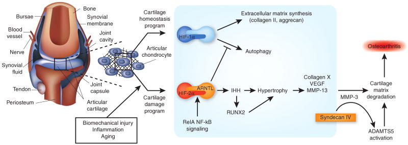
HIF-1α and HIF-2α have different functions in the cartilage. In the articular chondrocytes in the synovial joint, HIF-1α promotes homeostatic pathways, and HIF-2α promotes degradative pathways that foster osteoarthritis. The articular cartilage resides in hypoxic, avascular conditions within the synovial joint (left). Chondrocytes, cells of the articular cartilage, are affected by various forms of stress (biomechanical, inflammatory and aging), as well as the loss of synovial fluid boundary lubricants and the increase of certain factors released from subchondral bone and synovium. Four studies published in Nature Medicine have provided details for the emerging chondrocyte-centered osteoarthritis model depicted here–. Within the normal, unstressed chrondocyte (HIF-1α pathway), the hypoxic response transcription factor HIF-1α supports normal cartilage extracellular matrix synthesis and chondrocyte differentiation and promotes autophagy—all central activities in articular cartilage homeostasis. These effects of HIF-1α are antagonized by the closely related HIF-2α (HIF-2α pathway). HIF-2α promotes chondrocyte hypertrophy, a terminal differentiation state characterized by a unique gene expression program, including type X collagen and the type II collagen–degrading protease MMP-13. This switch to hypertrophy seems to be a relatively early signal to ignite and drive osteoarthritis in stressed cartilage. The preferential heterodimerization of HIF-2α with ARNTL creates the most potent set of partners for inducing chondrocyte hypertrophy. HIF-2α promotes hypertrophy, and complementary mechanisms (via IHH and RUNX2) stimulate increased expression of the major matrix-degrading protease ADAMTS5. The transmembrane heparan sulfate proteoglycan syndecan-4 (acting in part by inducing MMP-3 expression) stimulates activation of ADAMTS5 in hypertrophic chondrocytes. Inhibitors of ADAMTS5 and MMP-13 are already in clinical development. The model depicted here opens the door to additional, compelling strategies for potential disease modification in human osteoarthritis, such as suppressing the activity of HIF-2α, inhibiting the development of articular chondrocyte hypertrophy or targeting syndecan-4.
Comment on
-
Hypoxia-inducible factor-2alpha is a catabolic regulator of osteoarthritic cartilage destruction.Nat Med. 2010 Jun;16(6):687-93. doi: 10.1038/nm.2153. Epub 2010 May 23. Nat Med. 2010. PMID: 20495569
-
Transcriptional regulation of endochondral ossification by HIF-2alpha during skeletal growth and osteoarthritis development.Nat Med. 2010 Jun;16(6):678-86. doi: 10.1038/nm.2146. Epub 2010 May 23. Nat Med. 2010. PMID: 20495570
References
Publication types
MeSH terms
Substances
Grants and funding
LinkOut - more resources
Full Text Sources
Medical
