. 2010 Jul;12(7):703-10.
doi: 10.1038/ncb2073. Epub 2010 Jun 6.

Ciliary entry of the kinesin-2 motor KIF17 is regulated by importin-beta2 and RanGTP

Affiliations

Ciliary entry of the kinesin-2 motor KIF17 is regulated by importin-beta2 and RanGTP

John F Dishinger et al. Nat Cell Biol. 2010 Jul.

Abstract

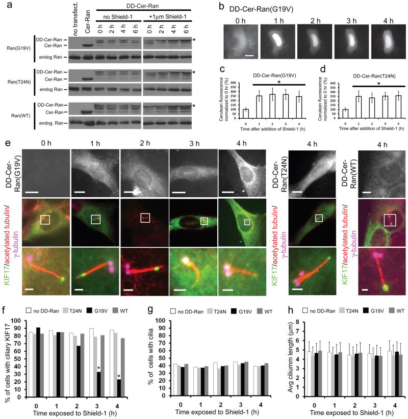
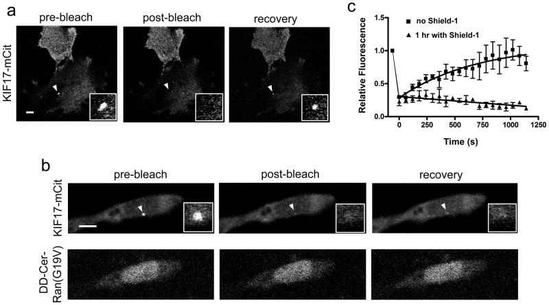
The biogenesis, maintenance and function of primary cilia are controlled through intraflagellar transport (IFT) driven by two kinesin-2 family members, the heterotrimeric KIF3A/KIF3B/KAP complex and the homodimeric KIF17 motor. How these motors and their cargoes gain access to the ciliary compartment is poorly understood. Here, we identify a ciliary localization signal (CLS) in the KIF17 tail domain that is necessary and sufficient for ciliary targeting. Similarities between the CLS and classic nuclear localization signals (NLSs) suggest that similar mechanisms regulate nuclear and ciliary import. We hypothesize that ciliary targeting of KIF17 is regulated by a ciliary-cytoplasmic gradient of the small GTPase Ran, with high levels of GTP-bound Ran (RanGTP) in the cilium. Consistent with this, cytoplasmic expression of GTP-locked Ran(G19V) disrupts the gradient and abolishes ciliary entry of KIF17. Furthermore, KIF17 interacts with the nuclear import protein importin-beta2 in a manner dependent on the CLS and inhibited by RanGTP. We propose that Ran has a global role in regulating cellular compartmentalization by controlling the shuttling of cytoplasmic proteins into nuclear and ciliary compartments.


Conflict of interest statement

COMPETING FINANCIAL INTEREST

The authors declare no competing financial interests.

Figures

Figure 1
Figure 1
The KIF17 CLS is necessary and sufficient for ciliary localization. (a) Odora, MDCK II, NIH3T3, and hTERT-RPE cells expressing full length KIF17-mCit (green) were fixed and stained for acetylated tubulin to mark cilia (red). Top row, images of entire cells; bottom row, higher magnification of cilia in boxed areas. White arrowheads indicate distal tips of cilia. (b) Left, schematic of full length human KIF17. NC, neck coil; CC, coiled-coil. Right, NIH3T3 cells expressing KIF17-mCit (green) were fixed and stained for acetylated tubulin (red) to mark cilia and γ-tubulin (blue) to mark the basal body. (c-g) Schematics of truncated and mutant KIF17 constructs (left) and their localization in Odora cells (right). Cells expressing the indicated truncated or mutant KIF17 motors (green) were fixed and stained for acetylated tubulin (red in c,f,g) or the myc tag (white in d). (h) Odora cells expressing full length KHC-mCit or KHC fused with the wildtype or mutant versions of the KIF17 tail were fixed and stained with antibodies to acetylated tubulin (red). Scale bars throughout figure are either 10 μm for images of entire cell or 1 μm for cilia.
Figure 2
Figure 2
Ran is present in the ciliary compartment. (a) Primary cilia were isolated from rat olfactory epithelium and the presence of Ran and adenylyl cyclase III (ACIII) in the ciliary (Cilia) and remaining deciliated (Decil.) fractions were determined by western blotting. Approximate molecular weights shown on left (kDa). Arrowheads designate protein of interest. Uncropped images of western blots are shown in Supplementary Fig. S7. (b) Representative compressed confocal stacks of coronal sections of rat nasal epithelia. Olfactory epithelia were immunostained with antibodies directed against Ran (left, green) and acetylated tubulin (middle, red). Merged imaged with Differential Interference Contrast (DIC) image is shown on right. Scale bar, 20 μm. Brackets denote cilia layer. Control images without primary antibody are shown in second row. (c) Immunofluorescence image of an NIH3T3 cell stained with anti-acetylated tubulin antibody (red), anti-RanGTP antibody (green), and DAPI (blue). Scale bar, 10 μm.
Figure 3
Figure 3
Fast upregulation of cytosolic Ran-GTP levels abolishes ciliary localization of KIF17. (a) COS cells expressing Cer-Ran (G19V, T24N, and WT), DD-Cer-Ran(G19V, T24N, or WT) or untransfected control cells were exposed to Shield-1 for 0 – 8 h (right panels) or untreated (left panels). The expression of endogenous and expressed Ran was determined by immunoblotting with an anti-Ran antibody. Uncropped images of western blots are shown in Supplementary Fig. S7. (b-d) Single cell analysis of live NIH3T3 cells expressing DD-Cer-Ran constructs upon exposure to Shield-1. (b) Representative images of a single cell expressing DD-Cer-Ran(G19V) at 0–4 h of Shield-1 exposure. Scale bar, 10 μm. The fluorescence increase in multiple cells was quantified for (c) DD-Cer-Ran(G19V) expressing cells (n=8) and (d) DD-Cer-Ran(T24N) expressing cells (n=10). Quantification includes both nuclear and cytoplasmic fluorescence. *, p<0.05 as compared to 0 h time point (two-tailed Student’s t-test). Data are presented as mean ± SEM. (e) NIH3T3 cells coexpressing KIF17-mCit and DD-Cer-Ran(WT) (right), DD-Cer-Ran(G19V) (left), or DD-Cer-Ran(T24N) (middle) were exposed to Shield-1 for 0–4 h and then fixed and stained for acetylated tubulin and γ-tubulin. (f-h) Quantification of the results in (e) to determine (f) the percentage of transfected cells with ciliary localization of KIF17-mCit (n=30 for each time point, collected over 3 experiments), (g) the percentage of transfected cells with cilia (n=50 for each), and (h) the cilium length in transfected cells (n=30 for each). *, p<0.05 as compared to 0 h of Shield-1 (Fisher’s Exact test). Data are presented as mean ± SD.
Figure 4
Figure 4
Upregulation of cytosolic Ran-GTP levels prevents ciliary entry of KIF17. FRAP analysis of Odora cells coexpressing KIF17-mCit and DD-Cer-Ran(G19V) in the (a) absence or (b) presence of Shield-1. The cells were imaged (pre-bleach) and then the fluorescence in the distal tip of the cilium was bleached at high laser power. The cells were again imaged (post-bleach) and the fluorescence recovery of KIF17-mCit in the cilium was monitored over time. White arrowheads, distal tips of cilia. Inset of each image is a close up of the cilium tip. Scale bars are 10 μm. (c) The fluorescence recovery of KIF17-mCit in the distal tips of cilia in the absence and presence of Shield-1 was quantified. Data are presented as mean ± SEM. n=5 for both traces. Data were subjected to single exponential curve fits.
Figure 5
Figure 5
KIF17 forms a complex with importin-β2 that is CLS- and Ran-GTP-dependent. (a) Odora cells were fixed and stained with antibodies to importin-β2 and acetelyated tubulin. Top row, images of entire cells; scale bar, 10 μm. Bottom row, magnification of boxed region containing cilia; scale bar, 1 μm. (b) Lysates of HEK293T cells expressing Flag-KIF17 or Flag-KIF17(1016-1019ala) or untransfected cells were immunoprecipitated with an anti-Flag antibody. The presence of expressed Flag-KIF17 and endogenous importin-β1 and -β2 proteins in the precipitates was probed by immunoblotting. (c) Lysates of HEK293T cells expressing Flag-KIF17 were immunoprecipitated with an anti-FLAG antibody in the absence (no Ran) or presence of the indicated purified GST-Ran proteins. The presence of expressed Flag-KIF17 and endogenous importin-β2 proteins in the precipitates was detected by immunoblotting. Uncropped images of western blots are shown in Supplementary Fig. S7. (d) (Left) Schematic of KIF17-mCit constructs in which the CLS is replaced with the NLS from SV40 T-antigen or the M9 NLS from hnRNP A1. (Right) Images of Odora cells expressing KIF17-mCit SV40 or M9 constructs and stained with anti-acetylated tubulin antibody. Scale bar, 1 or 10 μm. (e) Model for ciliary import of KIF17. In the cytoplasm (e′), KIF17 interacts with importin-β2 in a manner dependent on the KIF17 CLS. The importin/KIF17 complex is able to shuttle across the ciliary transition zone (e″) and into the cilium. Once across the barrier (e‴), the high levels of Ran-GTP in the cilium cause a dissociation of the KIF17-importin-β2 complex, allowing KIF17 to proceed with its role in IFT. Brown shading, subcellular areas of high RanGTP. Blue shading, subcellular areas of high Ran-GDP.

Comment in

Similar articles

Cited by

References

REFERENCES (MAIN TEXT)

    1. Silverman MA, Leroux MR. Intraflagellar transport and the generation of dynamic, structurally and functionally diverse cilia. Trends Cell Biol. 2009;19:306–316. - PubMed
    1. Scholey JM. Intraflagellar transport motors in cilia: moving along the cell’s antenna. J Cell Biol. 2008;180:23–29. - PMC - PubMed
    1. Satir P, Mitchell DR, Jekely G. How Did the Cilium Evolve? Ciliary Function in Mammalian Development. 2008;85:63–82. - PubMed
    1. Gerdes JM, Davis EE, Katsanis N. The Vertebrate Primary Cilium in Development, Homeostasis, and Disease. Cell. 2009;137:32–45. - PMC - PubMed
    1. Satir P, Christensen ST. Overview of structure and function of mammalian cilia. Annu Rev Physiol. 2007;69:377–400. - PubMed

REFERENCES (METHODS SECTION)

    1. Cai DW, Hoppe AD, Swanson JA, Verhey KJ. Kinesin-1 structural organization and conformational changes revealed by FRET stoichiometry in live cells. J Cell Biol. 2007;176:51–63. - PMC - PubMed
    1. Mayer U, et al. Proteomic analysis of a membrane preparation from rat olfactory sensory cilia. Chem Senses. 2008;33:145–162. - PubMed
    1. Mayer U, et al. The proteome of rat olfactory sensory cilia. Proteomics. 2009;9:322–334. - PubMed
    1. Davies S, Forge A. Preparation of the Mammalian Organ of Corti for Scanning Electron-Microscopy. J Microsc-Oxford. 1987;147:89–101. - PubMed

Publication types