. 2010 Jul;12(7):676-85.
doi: 10.1038/ncb2070. Epub 2010 Jun 6.

The matricellular protein CCN1 induces fibroblast senescence and restricts fibrosis in cutaneous wound healing

Affiliations

The matricellular protein CCN1 induces fibroblast senescence and restricts fibrosis in cutaneous wound healing

Joon-Il Jun et al. Nat Cell Biol. 2010 Jul.

Erratum in

  • Nat Cell Biol. 2010 Dec;12(12):1249

Abstract

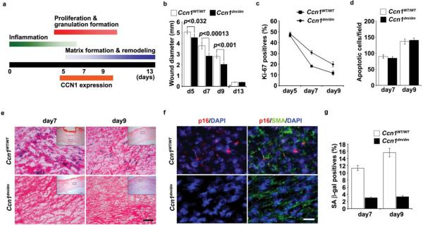
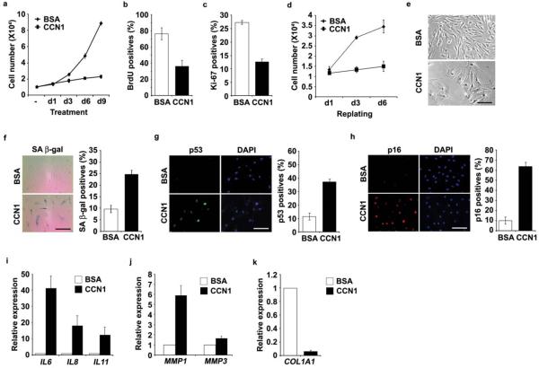
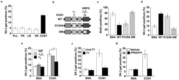
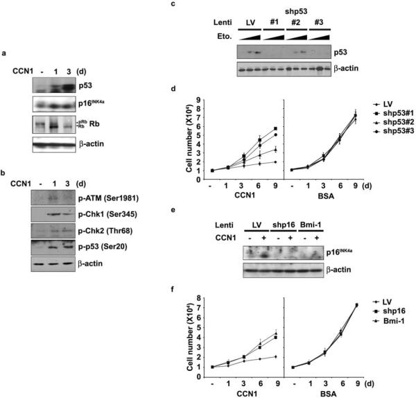
Cellular senescence is a recognized mechanism of tumour suppression; however, its contribution to other pathologies is not well understood. We show that the matricellular protein CCN1 (also known as CYR61; cysteine-rich protein 61), which is dynamically expressed at sites of wound repair, can induce fibroblast senescence by binding to integrin alpha(6)beta(1) and the heparan sulphate proteoglycans (receptors involved in cell adhesion). CCN1 induces DNA damage response pathways and activates p53 and the RAC1-NOX1 complex, which generates reactive oxygen species (ROS). This results in the ROS-dependent activation of the p16(INK4a)/pRb pathway, leading to senescence and concomitant expression of antifibrotic genes. Senescent fibroblasts accumulate in granulation tissues of healing cutaneous wounds and express antifibrotic genes in wild-type mice. These processes are lost in knockin mice that express a senescence-defective Ccn1 mutant, resulting in exacerbated fibrosis. Topical application of CCN1 protein to wounds reverses these defects. Thus, fibroblast senescence is a CCN1-dependent wound healing response in cutaneous injury that functions to curb fibrosis during tissue repair.


Figures

Figure 1
Figure 1. Impaired accumulation of senescent cells in Ccn1dm/dm mice during cutaneous wound healing
Excisional cutaneous wounds were created using a 6 mm biopsy punch in Ccn1WT/WT (n=5) and Ccn1dm/dm mice (n=5). (a) Time course of Ccn1 expression and overlapping phases of healing events. (b) Wound closure rate was monitored by measuring wound diameters on indicated days post-wounding. (c) The numbers of Ki-67-positive cells were counted as a percentage of Hematoxylin-positive nuclei in 5 randomly selected high-powered fields in frozen sections of granulation tissues, and (d) the numbers of apoptotic cells were counted after TUNEL staining. (e) Granulation tissues 7 and 9 days post-wounding were stained for SA-β-gal activity, and counterstained with Eosin. Inserts show the entire wound sections and the boxes indicate the enlarged areas. SA-β-gal positive cells were prominent in WT wounds but virtually undetectable in Ccn1dm/dm wounds. Similar results were observed in another independently-derived Ccn1dm/dm knockin mouse line. Scale bar = 50 μm. (f) Granulation tissues 9 days post-wounding were double-stained for p16INK4a (red) and α-SMA (green) by immunofluorescence, and counterstained with DAPI (blue). (g) Cells isolated from 7- and 9-day wounds were plated in 60 mm dishes and stained for SA-β-gal activity 2 days later (n=3).
Figure 2
Figure 2. CCN1 induces senescence in normal human fibroblasts
Young human BJ fibroblasts were treated with purified recombinant CCN1 protein (2.5 μg/ml). (a) Cells grown in the presence of BSA or CCN1 for indicated days were counted using a hemocytometer. After indicated treatments for 3 days, cells were subjected to either BrdU incorporation assay (b) or immunostaining for Ki-67 (c). BrdU or Ki-67 positive cells were counted and expressed as percentages of total number of cells in 10 randomly selected fields. (d) Cells were treated with CCN1 or BSA for 6 days, harvested by trypsinization and replated in full growth media. Cell proliferation was monitored by counting cell numbers. (e) Morphology of cells treated with CCN1. (f) SA-β-gal assay was performed and representative photomicrograph of cells (left) and quantification (right) are shown. Cells were subjected to immunofluorescence staining for p53 (g) or p16INK4a (h), and DAPI was used for counterstaining. Quantifications are shown on the right panels. (i–k) Cells were treated with CCN1 for 6 days and qRT-PCR was used to quantify the expression of proinflammatory cytokines (IL6, IL8, IL11), matrix degrading enzymes (MMP1, MMP3), and collagen 1 (COL1A1). Data represent mean ± S.D. of triplicate experiments. Scale bar = 100 μm.
Figure 3
Figure 3. CCN1 induces senescence through integrin α6β1-HSPGs
(a) Cells were adhered to dishes coated with ECM proteins including fibronectin (FN; 10 μg/ml), vitronectin (VN; 5 μg/ml), laminin (LN; 1 μg/ml), CCN1 (5 μg/ml) and poly-L-Lysine (PLL; 10 μg/ml), and stained for SA-β-gal after 3 days. (b) Schematic diagram of CCN1 showing the domain structure and the D125A and DM mutants, which are disrupted in binding sites for αv and α6β1-HSPG, respectively. (c) Cells were treated with either WT or mutant CCN1 proteins (2.5 μg/ml each) for 3 days and subjected to BrdU incorporation assay, and (d), SA-β-gal assay. (e) Cells were pre-incubated with function-blocking mAbs (50 μg/ml) against αvβ3 (LM609) or α6 (GoH3), and assayed for SA-β-gal. (f) Cells were treated with CCN1 with either the α6β1-binding T1 peptide or the non-binding mutant (mut-T1; 0.5 mM each), as a competitor, and SA-β-gal measured. (g) soluble heparin (1 mg/ml) was added 1 h before CCN1 treatment, and SA-β-gal assayed. Experiments were done in triplicates and data presented as means ± S.D. (*p<0.004).
Figure 4
Figure 4. Both p53 and p16INK4a participate in CCN1-induced senescence
(a) BJ cells were treated with CCN1 for indicated times, and expression of p53, p16INK4a, pRb, and β-actin was analyzed by immunoblotting. (b) Proteins involved in DDR, including phosphorylated forms of ATM (Ser1981), Chk1 (Ser345), Chk2 (Thr68), and p53 (Ser20) were detected by immunoblotting. (c) Cells were infected with three independent lentiviruses driving shRNAs against p53 (#1, 2 and 3). The effectiveness of knockdown was shown in cells treated with etoposide for 6 hrs (Eto; 20 or 50 μM) to induce p53. Cells infected with empty lentivirus (LV) were used as controls. (d) Cells infected as above were either treated with CCN1 or BSA and cell numbers were counted at indicated days. (e) Cells were infected with lentivirus expressing Bmi-1 or shRNA against p16INK4a, and (f) were either treated with CCN1 or BSA and cell numbers counted. Experiments were done in triplicates and data presented as means ± S.D.
Figure 5
Figure 5. Sustained ROS accumulation induced through CCN1-α6β1 interaction is required for senescence
(a) BJ cells were pretreated with NAC (2.5 mM) for 1 h followed by the addition of CCN1. NAC was replenished daily and cell numbers were counted, and (b) SA-β-gal expression was evaluated after 3 days. (c) Cells were pre-treated with function-blocking mAbs against αvβ3 or α6 (50 μg/ml each) for 1 h, followed by CCN1 treatment for an additional 1 h and stained with H2DCFDA (10 μM). ROS was quantified by fluorescence measurements. (d) Cells were adhered to surfaces coated with PLL or ECM proteins including FN, VN, LN, Collagen I (Col1; 10 μg/ml), and CCN1 as in Fig. 3a, and ROS levels were measured after 1 or 4 h as above. (e) Cells were treated with CCN1 and SA β-gal expression was assayed after 3 days. NAC (2.5 mM) was added either 1 h before or 3, 6, 10, and 24 h after CCN1 treatment. All experiments were done in triplicates and data presented as means ± S.D.
Figure 6
Figure 6. NOX1-RAC1 complex mediates CCN1-induced senescence
(a) BJ cells were preincubated for 1 h with inhibitors of 5-LOX (MK886, 5 μM), NADPH oxidase (apocynin, 10 μM) or vehicle (0.1% DMSO), followed by CCN1 treatment for 3 days and SA-β-gal expression was determined. Chemical inhibitors were replenished daily. (b) Cells were infected with lentiviruses encoding shRNAs against 5-LOX (sh5LOX #1 and #2). Knockdown was assessed by immunoblotting (left) and cell proliferation after CCN1 treatment was monitored by counting cell numbers (right). (c) Cells treated with CCN1 for various times were assayed for expression of NOX1, NOX4, catalase and β-actin by RT-PCR. (d) Lentiviral shRNA-mediated knockdown of NADPH oxidase 1 (shNOX1 #1 and #2) and 4 (shNOX4) was accomplished as above and confirmed by RT-PCR (top). Proliferation of infected cells after CCN1 treatment was determined by counting cell numbers (bottom). (e) Lentiviral shRNA knockdown of RAC1 (shRAC1) was confirmed by immunoblotting (top), and proliferation of knockdown cells after CCN1 treatment was assessed (bottom). All experiments were done in triplicates and data presented as means ± S.D.
Figure 7
Figure 7. Induction of p16INK4a through ROS-dependent activation of stress kinases
(a) Lysates of BJ cells treated with CCN1 for indicated times were analyzed for the activation of ERK1/2 (p42/p44), p38 MAPK, and JNK1/2 by immunoblotting using their cognate antibodies and phosho-specific antibodies. (b) Cells were pre-incubated with inhibitors of either p38MAPK (SBS202190; 10 μM), MEK1 (PD98059; 20 μM), or JNK1/2 (SP600125; 25 μM) for 1 h and then treated with CCN1 (2.5 μg/ml). SA-β-gal expression was assayed three days later. Data presented as means ± S.D. (n=5) (c). Cells pre-incubated with inhibitors as above were then treated with CCN1 for 24 or 48 hrs, and p16INK4a was detected by immunoblotting. (d) Activation of both ERK and p38 MAPK by CCN1 was examined in cells pretreated with either NAC (2.5 mM) or apocynin (10 μM) by immunoblotting. (e) A signaling model for CCN1-induced senescence. Upon binding to integrin α6β1 and HSPGs, CCN1 activates the RAC1-NOX1 complex to generate a robust and sustained accumulation of ROS, which triggers the biphasic hyperactivation of ERK and p38 MAPK, leading to p16INK4a induction. CCN1 also induces a DDR and activates p53, in part through a ROS-dependent mechanism. Both p53 and p16INK4a contribute to CCN1-induced senescence.
Figure 8
Figure 8. Enhanced fibrogenic response during wound healing in Ccn1dm/dm mice
Gene expression in granulation tissues from WT and Ccn1dm/dm mice 5–13 days post-wounding was analyzed by qRT-PCR. Relative expression of (a) Mmp2, Mmp3, Mmp9, and (b) Col1a1 and Tgfβ1 is shown. (c) Total amount of hydroxyproline in healing wounds isolated 5–21 days post-wounding from WT and Ccn1dm/dm mice was determined and normalized over the total dry weight of the tissue. (d) Frozen sections from tissues 9 days post-wounding were stained with Sirius red for collagen. Images were acquired from bright field (top) and under polarized light (bottom). Scale bar = 100 μm. (e) Sirius red-positive area was calculated from at least 3 adjacent sections using ImageJ software. All experiments were done in triplicates and data presented as means ± S.D. (f) Purified recombinant CCN1 protein (0.1 mg/ml; 50 μl per dose) in PBS or PBS alone was topically applied to excisional wounds of Ccn1dm/dm mice daily, and wound granulation tissue harvested 9 days post wounding and analyzed by qRT-PCR (n=5). (g) Wounds so treated were sectioned and stained with Sirius red and visualized under polarized light (scale bar = 100 μm), and (h) stained for SA-β-gal (scale bar = 50 μm).

Similar articles

Cited by

References

    1. Hayflick L. The limited in vitro lifetime of human diploid cell strains. Exp. Cell Res. 1965;37:614–636. - PubMed
    1. Campisi J, d'Adda di Fagagna F. Cellular senescence: when bad things happen to good cells. Nat. Rev. Mol. Cell Biol. 2007;8:729–740. - PubMed
    1. Collado M, Blasco MA, Serrano M. Cellular senescence in cancer and aging. Cell. 2007;130:223–233. - PubMed
    1. Braig M, et al. Oncogene-induced senescence as an initial barrier in lymphoma development. Nature. 2005;436:660–665. - PubMed
    1. Chen Z, et al. Crucial role of p53-dependent cellular senescence in suppression of Pten-deficient tumorigenesis. Nature. 2005;436:725–730. - PMC - PubMed

Publication types