

doi: 10.1038/nsmb.1852.
Epub 2010 Jun 6.
Mutagenic conformation of 8-oxo-7,8-dihydro-2'-dGTP in the confines of a DNA polymerase active site
Affiliations
- PMID: 20526335
- PMCID: PMC2921931
- DOI: 10.1038/nsmb.1852
Item in Clipboard
Mutagenic conformation of 8-oxo-7,8-dihydro-2'-dGTP in the confines of a DNA polymerase active site
Nat Struct Mol Biol.
2010 Jul.
Abstract
The major product of oxidative base damage is 8-oxo-7,8-dihydro-2'-deoxyguanine (8odG). The coding potential of this lesion is modulated by its glycosidic torsion angle that controls whether its Watson-Crick or Hoogsteen edge is used for base pairing. The 2.0-A structure of DNA polymerase (pol) beta bound with 8odGTP opposite template adenine indicates that the modified nucleotide assumes the mutagenic syn conformation and that the nonmutagenic anti conformation would be incompatible with efficient DNA synthesis.
Figures
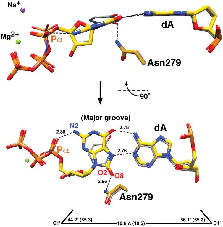
Syn-conformation of 8odGTP paired with adenine. The structure of the substrate complex of pol β with a correct incoming nucleotide (dUMPNPP; PDB ID 2FMQ, gray carbons) was superimposed with the structure with an incoming 8odGTP paired with adenine (yellow carbons). Two views of the nascent base pair binding pocket illustrate the syn-conformation (χ = 51°) of 8odGTP forming a Hoogsteen base pair with adenine (dA). The geometry (C1′ distance and λ angles) of the mismatched base pair is shown at the bottom (values for a correct nascent base pair are in parentheses). The purple (Na+) and green (Mg2+) spheres represent modeled ions in the catalytic and nucleotide binding sites of the mismatched structure, respectively.
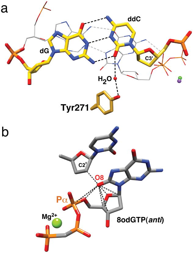
Structural features that discourage insertion of 8odGTP. (a) Although the 8odGTP(syn)—dA(anti) nascent base pair (gray carbons, wire representation) displays good geometry, the primer terminus base pair (yellow carbons) is displaced into the major groove in an attempt to maximize stacking interactions with 8odGTP(syn). This results in a loss of a direct hydrogen bond with Tyr271. Accordingly, the geometry between C3′ and Pα (8odGTP) is distorted. (b) Modeling a carbonyl at C8 of an incoming dGMPPCP(anti) paired with cytosine (PDB ID 2ISP) suggests that steric repulsion between the sugar-phosphate backbone and the sugar (C2′) of the primer terminus (dashed lines; distances tabulated in Supplementary Table 3) would reduce correct insertion of 8odGTP.
References
-
- Ames BN, Gold LS. Mutat Res. 1991;250:3–16. - PubMed
-
- Kouchakdjian M, et al. Biochemistry. 1991;30:1403–1412. - PubMed
-
- McAuley-Hecht KE, et al. Biochemistry. 1994;33:10266–10270. - PubMed
-
- Maki H, Sekiguchi M. Nature. 1992;355:273–275. - PubMed
-
- Beard WA, Wilson SH. Chem Rev. 2006;106:361–382. - PubMed
Publication types
MeSH terms
Substances
Associated data
- Actions
Grants and funding
LinkOut - more resources
Full Text Sources
Miscellaneous
